11. The Eye. Anatomy Of The Eye
Description
This section is from the book "The Horse - Its Treatment In Health And Disease", by J. Wortley Axe. Also available from Amazon: The Horse. Its Treatment In Health And Disease.
11. The Eye. Anatomy Of The Eye
The eye is an instrument by which light, colour, form, and movement are recognized, and by which, combined with other faculties, we acquire a knowledge of distance, relation, position, and size of objects.
It occupies the fore or outer part of the cavity named the orbit, the bones of which form a very efficient means of protection to it against injury. It rests on a soft bed of fat, which enables it to yield to direct blows, and it is further protected by a retractor muscle, which withdraws it under cover of the bones, whilst the eyelids and the membrana nictitans (haw) cover it in front. It is consequently only rarely injured. The membrana nictitans, or haw, is a triangular piece of cartilage which gradually becomes thinner from back to front. It is situated in the inner canthus of the eye, and is attached to a cushion of fat by its posterior angle, while in front it presents a very thin edge by which small particles of dust, hairs, insects, and the like, are removed. If not swept away by the nictitating membrane, they are washed away by the copious secretion of tears which they excite from the lachrymal gland (fig. 246, e).
It is moved or rotated by seven muscles, viz., the four recti, or straight muscles, named respectively the superior, inferior, external, and internal rectus; the superior and inferior oblique muscles, and the retractor bulbi. The superior oblique muscle, which turns the eye downwards and outwards, is supplied by the fourth nerve or trochlearis. The external rectus is supplied by the sixth cranial nerve, and the remaining five muscles are supplied by the third nerve. These muscles, with the exception of the inferior oblique, arise from the back of the orbit, and, passing forwards, are inserted into the fore part of the globe. The inferior oblique muscle arises from the inner part of the floor of the orbit, and. running outwards and upwards, forms a kind of sling - supporting the eye together with the tendon of the superior oblique. The superior oblique (fig. 239, e), arising from the back of the orbit, ends in a tendon which runs through a small loop or pulley on the upper part of the inner wall of the orbit, and then, changing its direction, runs outwards and a little backwards, to be inserted into the upper part of the globe.
The ancient division of the structures of the eye into three coats or tunics, and three humours, is still the most convenient for description. The first coat is composed of the sclerotic and cornea (fig. 240, D, l).
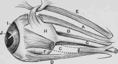
Fig. 239. - Muscles of the Eyeball.
A, Superior Rectus Muscle. B, Retractor Muscle. C, External Rectus Muscle. D, Inferior Rectus Muscle. E, Superior Oblique Muscle. F, Point of Insertion of Inferior Oblique Muscle. 0, Optic Nerve. H, Globe of the Eye. I, Iris. J, Pupil.

Fig. 240. - Section of Eye.
A, Lachrymal Gland. B, Levator Palpebrae Superioris. C, Levator Oculi. D, Sclerotic Coat. E, Choroid Coat. F, Retina. G, Optic Nerve. H, Vitreous Humour. I, Capsule of the Lens. J, Crystalline Lens. K. Aqueous Humour. L, Cornea. M, Iris. N, Upper Eyelid.
The second coat is formed by the choroid ciliary processes and iris (fig. 240, E, m).
The third coat is the retina, or expansion of the optic nerve (fig. 240, f). The three humours are the aqueous humour (fig. 240, k), the crystalline lens (fig. 240, j), and the vitreous humour (fig. 240, h).
The outermost tunic is a dense, tough, and unyielding membrane, the inner four-fifths of which is opaque, and named the sclerotic or white of the eye, whilst the outer or front fifth is transparent, and is named the cornea or glass of the eye. The sclerotic is thick behind, where it presents a small opening for the entrance of the optic nerve (fig. 248, 1), and thinner in front, where it becomes continuous with the cornea. The fore part of the sclerotic, or that which forms the white of the eye, is covered with a delicate mucous membrane, named the conjunctiva, which is continued over the cornea as a transparent membrane composed of eight or ten layers of cells. These cells, if injured, can be thrown off and renewed, so that the transparency of the cornea is not impaired after slight lesions. Different as the aspect of the cornea is from the white or sclerotic portion of the membrane, the material out of which each is composed - connective tissue - is the same, only that in the sclerotic the fibres are irregularly arranged, whilst in the cornea they are disposed in layers or lamellae one upon another, with many intervening branching cells which enable the nutrient fluid or plasma of the blood to penetrate and nourish the tissue. The cornea is supplied by the fifth pair of nerves, which confer upon it the exquisite sensitiveness of the surface.
The Choroid Tunic is composed of a close net-work of blood-vessels, the outer layer of which is formed by the large veins of the eye known as venae vorticosae (fig. 241, 2). The inner layer is formed by the delicate capillary vessels disposed in loops, and is in contact with the retina. At the back part of the choroid there is a circular opening, like that in the sclerotic, for the passage of the optic nerve to the retina; in front the choroid is folded into a large number of plaits named the ciliary processes (fig. 242, c), which project into the interior of the eye behind the iris.

Fig. 241. - The Choroid Tunic.
1, Cut Surface of Sclerotic. 2, Veins of the Choroid (Venae Vorticosae). 3, Ciliary Nerves and Arteries. 4, Ciliary Ligament. 5, Iris. 6, Pupil.
Numerous dark-looking pigment cells, of irregular forms, are distributed between the blood-vessels, giving to the membrane a soft, velvety-black colour, except at one part, which forms a broad band just above the entrance of the optic nerve. Here the choroid presents a metallic yellowish-green colour, and reflects the light strongly.
This appearance is attributed to the peculiar arrangement of some thin cell-plates found in this part, producing the phenomena of interference of light, and is believed to assist the animal in perceiving objects in twilight.
The Ciliary Processes (fig. 242, c), which are thickly covered with black pigment, are about one hundred and twenty in number, and are arranged in a circle, projecting into the interior of the globe to become connected with the vitreous humour. They probably play an important part in the secretion of the aqueous humour. Externally they present a whitish band, which is the ciliary body or muscle. Some of the fibres of this muscle radiate backwards from the margin of the cornea over the choroid, others are circular; both, when acting, modify the curvation of the lens, and thus influence the accommodation of the eye. The Iris (fig. 241, 5) is a thin membrane composed of blood-vessels and of muscular fibres united together by connective tissue. Some of these fibres are arranged in the form of a ring around the inner margin of the pupil, while a second set, outwardly placed to these, and connected with them, are disposed in a radiating manner like the spokes of a wheel. In the centre of the iris is an ovoid opening, the "pupil " (fig. 241, 6), on one or both edges of which are a few small black bodies called corpora nigra (fig. 242, d). The pupil is the opening through which light enters the eye, and by which images of objects in the outer world are permitted to be formed on the retina within. The iris, except in albinos, is perfectly opaque, and acts as a diaphragm, regulating the amount of light that enters the eye. In dim lights the pupil dilates (fig. 243), in bright lights it contracts (fig. 244).

Fig. 242. - Interior View of the Eye.
A, Pupil. B, Iris, c, Ciliary Processes. D, Corpora Nigra.

Fig. 243. - Pupil Dilated.

Fig. 244. - Pupil Contracted.
Continue to:
My Books

