Anatomy Of The Female Organs Of Generation
Description
This section is from the book "The Horse - Its Treatment In Health And Disease", by J. Wortley Axe. Also available from Amazon: The Horse. Its Treatment In Health And Disease.
Anatomy Of The Female Organs Of Generation
The entrance to the uro-genital passage in the female is a vertical fissure some 4 or 5 inches in length, bounded on each side by a skin fold (the labium magnus). The folds meet above at an acute angle, separated from the anus by a bare, usually dark-coloured patch of skin, about 2 inches in extent, known as the " perineum ". Below, the meeting of the lips is more obtuse. Supporting the folds, and passing round the opening, is a voluntary muscle (post sphincter), which is separated from the skin by a considerable quantity of loose areolar tissue.
If the labia are separated it will be seen that they are covered internally by a smooth, moist, pale-coloured mucous membrane. Just within the lips, in the lower angle of the fissure, will be noticed a small prominence something like the glans penis of the male. This is the "clitoris"; the little body is partly contained within a prepuce formed by the coming together of two short folds of mucous membrane corresponding with the " labia minora". The integument covering the organ and lining its sheath is richly supplied with glands of a sebaceous type, which form a substance similar to the blackish gray secretion (smegma) commonly found in the sheath of the male. The clitoris can be raised and its glans turned directly backwards. This action is well seen immediately after urination and at frequent intervals during the period of oestrum.
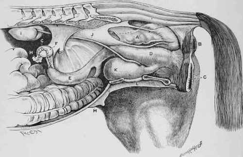
Fig. 228. - View of the Genitourinary Organs of the Mare.
A, Rectum. B, Anus. c, Vulva. D, Vagina. E, Left Horn of Uterus. F, Ovary. G, Fallopian Tube. H, Its Fimbriated extremity. I, Kidney. J, Ureter. K, Bladder. L, Pelvis. M, Mammary Gland.
The cavity between the labia, into which the hand can be introduced, is the "vulva"; it extends inwards for 4 or 5 inches, and is bounded in front by the rudiment of the hymen, a small membranous fold which separates it from the vagina. The vulva is lined by pink mucous membrane, and kept moist by the secretion from numerous small mucous glands embedded in its structure. Outside the mucous membrane there is much loose connective tissue to allow of expansion of the passage during parturition, and large plexuses of veins. Outside this again are muscle fibres continuous with the posterior sphincter, and which in front become thickened to form an anterior sphincter. Opening on to the floor of the cavity, about 4 inches within the lips, is the urethra or urinary passage (fig. 229, h). This orifice is covered by an ample fold of mucous membrane having its free edge directed backwards; above it is the large orifice leading to the vagina, encircled at its margin by the rudiment of the hymen, a membranous fold often hardly noticeable. When present in a state of full development it is ruptured at the first service, and its remains are then known as the "carunculae myrtiformes". The vagina extends forwards from the hymen to the uterus, and is about 9 inches in length. Projecting into its anterior extremity for about 1 1/2 inch is the neck of the uterus. The vaginal passage is lined by pale-red mucous membrane thrown into longitudinal ridges extending along its whole length. Outside the mucous membrane is a plentiful areolar tissue in which are large venous plexuses and layers of unstriated muscle fibre; outside this again are muscular layers, longitudinal and circular, continuous with those of the uterus. Beneath the vagina are the urethra and the bladder (k, fig. 228), and above it the rectum (a, fig. 228).
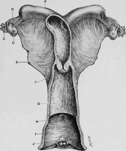
Fig. 229. - Female Organs of Generation.
A, Left Horn of Uterus. B, Fallopian Tube, c, Fimbriated extremity of the same. D, Ovary. E, Broad Ligament, F, Os Uteri. G, Vagina. H, Opening into Bladder. I, Vulva. J, Clitoris.
Continue to:
My Books

