Examination Of The Eyes
Description
This section is from the book "The Horse - Its Treatment In Health And Disease", by J. Wortley Axe. Also available from Amazon: The Horse. Its Treatment In Health And Disease.
Examination Of The Eyes
For the purpose of making an examination of the eye, the horse should be so placed by the observer that the light may fall on the organ either from a window or from the stable entrance, while the animal's head is either within the stable or only partly out of it. In this position the examiner will naturally first note the condition of the eyelids and the movements of the eye. If, during this time, the animal happens to be suddenly alarmed, even by the movement of a hand towards the eye, he will also witness the rapid protrusion of the haw (fig. 247) (cartilago nictitans) over the front of the organ, as if to protect it from injury. In the section on anatomy and physiology the structure and uses of this organ are described.
Having the eye in a good light, the examiner will note the white portion of the globe, with its numerous vessels ramifying in all directions, and the transparent cornea forming the front of the eye. Looking through the cornea he will observe the curtain behind it which gives the colour to the eye, and is therefore termed the iris, with the opening in the centre of it, the pupil, through which the light passes, carrying with it the image which is to be depicted on the retina. At the edge of the pupilary opening the small black, pea-like bodies, which have been described as the corpora nigra, will be seen; small, or even extremely minute, at the lower edge of the pupil, considerably larger at the upper edge, these little bodies are worth particular attention, because they are constantly taken by the inexperienced for a diseased condition of the eye. Looking through the pupilary opening, if the eye is in the proper position, the examiner will see a small pearl-like body at the bottom of the posterior chamber which marks the entrance of the optic nerve (fig. 248, l); this body is worthy of particular notice as it is frequently mistaken for a cataract.
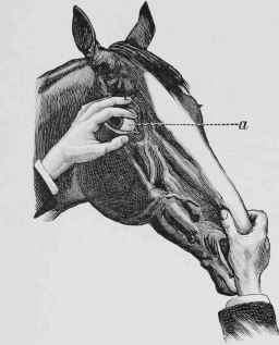
Fig. 247. - Examination of the Eye (a, the Haw).
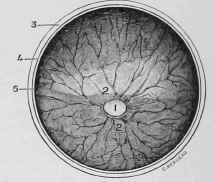
Fig. 248. - Fundus of the Eye.
1, Entrance of the Optic Nerve to form the Retina. 2, 2, Blood-vessels entering around it (there is no arteria centralis retinae in the horse as in man). 3, Divided Sclerotic. 4, Divided Choroid. 5, Divided Retina.
A quarter of an hour occupied in this examination every day for a week should make the tyro familiar with some of the more important parts of the anatomy of the visual organ and its appendages, so that he will be able to recognize any decided changes resulting from disease without running the risk of making the curious mistakes which are so extremely common.
During the examination the observer will note that the eye of the horse is never for more than a second in one position. In examining the eye of a friend this difficulty entirely vanishes, as the organ can be kept perfectly still while the inspection is being made, but there are positively no means of securing this fixity in the eye of the lower animals except by putting them under the influence of an anaesthetic, and anyone desiring to examine the eye is required to train his own eyes to follow the movements of the organ, so that he may keep the part which he wishes to see in view during the time that it is in constant motion.
Continue to:
My Books

