Loose Cartilages In Joints
Description
This section is from the book "The Horse - Its Treatment In Health And Disease", by J. Wortley Axe. Also available from Amazon: The Horse. Its Treatment In Health And Disease.
Loose Cartilages In Joints
It sometimes occurs that small bodies, varying in size from a pea to a walnut, are found loose in the cavities of joints, especially the larger ones, such as the stifle, hock, or knee.
These formations are generally ovoid in shape and somewhat flattened. In colour they are yellowish-gray or grayish-white, and vary in composition not only in different cases but in the same joint.
Some are composed of cartilage or fibro-cartilage, interspersed or not with bony matter, while a few are almost entirely made up of the last-named substance.
Some of these formations originate as outgrowths from the internal surface of the synovial membrane, from which they hang suspended for a time, and are then broken away by the movements of the joint and become free or, as they are termed, "loose cartilages".
Others more distinctly cartilaginous in type commence as small excrescences along the margin of the articular cartilage, and these, like those last referred to, are rubbed off, and when disconnected move about the joint, interfering with action and causing pain and lameness, which may be continuous or intermittent.
As to whether these excrescences grow after their detachment from their place of origin it would be difficult to say, but there is reason to think that such is sometimes the case.
Symptoms
While connected with the synovial membrane or the cartilage these growths may occasion very little disturbance, and even when detached, small ones, while in a soft, fibrous, or cartilaginous condition, do not seriously interfere with action; but the larger and harder ones provoke serious attacks of lameness by becoming fixed between the ends of the bones and the capsular membrane and otherwise damaging the joint.
In a case which occurred in a three-year-old colt under the care of the late Mr. Joseph Axe of Doncaster, and which the writer had an opportunity of seeing and examining after death, the patient was slightly lame of the near hind - leg for several weeks before the seat of lameness could be localized. Suddenly the animal became incapable of advancing the limb, and exhibited considerable pain when caused to move. The stifle-joint then began to swell owing to distension of the capsule with fluid; there was also considerable heat and tenderness to pressure. The swelling continued, but the lameness almost entirely disappeared in a week, only, however, to return with increased severity a fortnight later. This subsidence and return of the acute symptoms was repeated on three or four occasions, each time leaving the joint larger and the lameness more severe. Ultimately the colt was destroyed, and three loose cartilages (fig. 362), one being an inch and a half long and three-quarters of an inch wide, were removed from the joint capsule.
The two smaller ones consisted of fibro-cartilage, and the largest of cartilage interspersed with bony matter. The joint contained a large quantity of dark, straw-coloured fluid, and the synovial membrane was considerably inflamed and thickened.
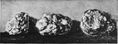
Fig. 362. - Loose Cartilages removed from the Capsule of a Stifle-joint.
Treatment
Nothing short of the removal of the offending bodies by a surgical operation can be of service in these cases, but the difficulty interposed to an accurate diagnosis by the thickness of the skin, the ligaments, and parts about the joint, renders such a course as unreliable as it is dangerous.
The remedy, indeed, may prove even worse than the disease.
Continue to:
My Books

