The Humours Of The Eye
Description
This section is from the book "The Horse - Its Treatment In Health And Disease", by J. Wortley Axe. Also available from Amazon: The Horse. Its Treatment In Health And Disease.
The Humours Of The Eye
The aqueous humour (fig. 240, k) is a limpid fluid which occupies the space between the cornea and the lens. The quantity is estimated at about 1 fluid drachm. It appears to be secreted by the ciliary processes, and undergoes constant renewal. That which is freshly secreted is poured forth into the posterior chamber of the eye, and therefore occupies the space between the back of the iris, in front, and the tips of the ciliary processes, the suspensory ligament of the lens, and the lens itself, behind. The fluid passes through the aperture of the pupil into the anterior chamber of the eye, and escapes by a natural channel situated in the angle of this chamber at the line where the iris and cornea are in contact. The channel is named the canal of Fontana, and communicates with another canal named the canal of Schlemm.
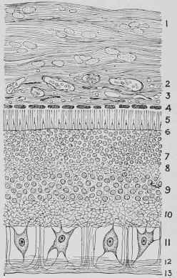
Fig. 245. - Microscopic Section of the Retina, Choroid, and Part of the Sclerotic.
1, Sclerotic. 2, External Vascular Portion of the Choroid. 3, Internal Vascular Portion of the Choroid. 4, Pigment Cell Layer. 5, Layer of Rods and Cones. (5, Membrana Limitans Externa. 7, External Nuclear Layer. 8, External Molecular Layer. 9, Internal Nu-clear Layer. 10, Internal Molecular Layer. 11, Ganglion Layer. 12, Nerve-fibre Layer traversed by Mutter's Sustentacular Fibres. 13, Membrana Limitans Interna.
The Lens (fig. 240, j) is a perfectly transparent biconvex solid body, about half an inch in diameter. The front surface is less convex than the posterior. It lies behind the iris, and is lodged in a depression of the vitreous humour. It is kept in position by a circular ligament which is formed out of a continuation of the modified outer layer of the retina and extends from the ciliary processes to the margin of the lens.
The lens is enclosed in a capsule, which is thick in front and thin behind. It is composed of long fibres with sawlike edges, which mutually interlock with those of the adjoining fibres; and these are arranged in many layers, the outermost being soft, becoming denser as the central part of the lens is reached. By the lens, parallel or nearly parallel rays of light are brought to a focus on the retina, and a precise image is there formed. Opacity of the lens constitutes cataract.
The Vitreous Humour (fig. 240, h) is a semi-fluid, uniform substance, like white-of-egg in consistence, traversed by numerous delicate fibres which appear to be the remains of cells with greatly attenuated and elongated processes. It is enclosed in a transparent and very thin membrane termed the hyaloid membrane. The vitreous humour presents a canal running through its centre from behind, which originally contained blood-vessels on their way to the posterior surface of the lens, but both the canal and the blood-vessels disappear in infancy. In front the vitreous humour is hollowed out, so as to give lodgment to the convex surface of the lens in front of it. Near the margin of the lens' is a structure named the Zonule of Tinn, composed of fibres which are superimposed upon the hyaloid membrane and forming the suspensory ligament of the lens, whilst they are so arranged as to present alternate elevations and depressions which correspond to the ciliary processes and the intervening spaces.

Fig. 246. - The Eye, showing the Lachrymal Gland.
a, Superior Oblique Muscle. B, Levator Oculi or Superior Rectus, c, Abductor Oculi or External Rectus. D, Depressor Oculi or Inferior Rectus. E, Lachrymal Gland. F, Optic Nerve.
Continue to:
My Books

