


The Veins. Continued
Description
This section is from the book "Handbook Of Anatomy For Students Of Massage", by Margaret E. Bjorkegren. Also available from Amazon: Handbook Of Anatomy For Students Of Massage.
The Veins. Continued
Jugular Veins
Common facial, formed by union of facial and a terminal branch of the temporo-maxillary veins, which drain the muscles of the face and fore-part of the scalp.
Occipital vein, which drains the back part of the scalp.
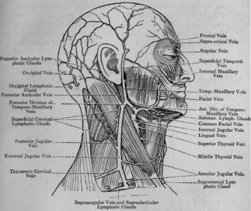
Fig. 59. - Veins and Glands of Head and Neck.
The Subclavian vein is the direct continuation of the axillary vein. It commences at the lower border of the first rib and passes in front of scalenus anticus, which separates it from the artery, and terminates behind the sternal end of the clavicle by uniting with the internal jugular vein to form the innominate vein.
Tributary
External jugular formed on the surface of the sterno-mastoid by the union of the terminal branch of the temporo-maxillary with the posterior auricular vein, which drain the outer side of the head and neck.
The Inferior Vena Cava is formed opposite the right side of the body of the fifth lumbar vertebra behind and external to the right common iliac artery by the union of the common iliac veins. It passes up the posterior wall of the abdomen to the right of the aorta and on the right crus of the diaphragm, and passes through the latter at the level of the eighth dorsal vertebra. It then pierces the pericardium and enters the lower and back part of the right auricle. The vessel lies below on the bodies of the lumbar vertebrae, the right psoas, and the right crus of the diaphragm, the right renal artery, and suprarenal capsule. Anterior to it are the right common iliac artery, the third part of the duodenum, head of the pancreas, the portal vein, the first part of the duodenum, and the posterior surface of the liver. On its left side are the aorta and the left crus of the diaphragm.
Tributaries
Hepatic.
Inferior phrenic.
Suprarenal.
Renal.
Lumbar.
Spermatic or ovarian.
The Hepatic veins are two in number, which open into the inferior vena cava just below the diaphragm, and bring the blood from the liver which has entered it through the hepatic artery and portal vein.
The Inferior Phrenic veins are formed by the venae comites of the arteries supplying the diaphragm.
The Suprarenal veins are one each from the suprarenal capsules. Sometimes the one on the left side enters the left renal vein.
The Renal veins each issue from the hilum of the kidney, the left one being longer than the right. The left one crosses in front of the left psoas, the left crus of the diaphragm, and the aorta just below the superior mesenteric artery, and lies behind the pancreas and the last part of the duodenum. The right renal vein passes behind the duodenum.
The Lumbar veins, of which there are four pairs, are formed by tributaries from the lateral and posterior walls of the abdomen. They are also connected together by the azygos veins, anastomosing vessels passing upwards.
The Spermatic or Ovarian veins are formed by the pampiniform plexuses surrounding the testicles or ovaries. The vein on the right side enters the inferior vena cava, that on the left usually enters the left renal vein.
The Common Iliac veins are formed by the union of the external iliac and internal iliac veins opposite the brim of the pelvis behind the internal iliac artery. They pass upwards and inwards, and unite to form the inferior vena cava opposite the fifth lumbar vertebra.
Tributaries
External iliac. Internal iliac.
Ilio-lumbar, small veins from the iliac fossa and adjacent parts.
The Internal Iliac vein is formed at the upper border of the great sciatic notch by the union of veins corresponding to the branches of the internal iliac artery, except the ilio-lumbar branches. It lies behind and slightly to the inner side of the corresponding artery.
The External Iliac vein is the continuation of the femoral vein, and commences on the inner side of the femoral artery. Passing upwards and inwards along the brim of the pelvis, it ends by joining the internal iliac to form the common iliac vein.
The Portal System
The portal vein is formed by tributaries from the whole of the alimentary canal (except the lower part of the rectum), the spleen and the pancreas, and conveys the blood thus collected to the liver. The tributary veins correspond at first with the arteries supplying these organs, but the terminal veins differ somewhat. The inferior mesenteric vein joins the splenic vein, and they join with the superior mesentric vein to form the portal vein. The portal vein is a short wide vessel about three inches long, which commences behind the neck of the pancreas in front of the left border of the inferior vena cava. It passes upwards behind the pancreas, duodenum, and common bile-duct, and enters the transverse fissure of the liver. The Middle and Inferior Haemorrphoidal veins which drain the lower part of the rectum, enter the internal iliac vein, and thus connect the vena cava and portal systems.
Continue to:


My Books

