


The Head, Face, And Neck
Description
This section is from the book "Lessons on Massage", by Margaret D. Palmer. Also available from Amazon: The Book Of Massage: The Complete Stepbystep Guide To Eastern And Western Technique.
The Head, Face, And Neck
The bones of the head and face are called the skull. It is divided into the cranium and face. In the cranium are eight bones ;
1 Frontal (forehead). 2 Parietal (sides). 2 Temporal (temples)
1 Occipital (back of head). 1 Ethmoid (sieve). 1 Sphenoid (wedge).

Fig. 89.—1, Frontal bone ; 2, occipital bone ; 3, upper maxillary ; 4, lower maxillary; 5, mastoid process on temporal bone.
The frontal bone is the forehead. It forms the upper and fore part of the head, and upper part of the socket of the eye.
The parietal are the side walls, which unite and form the arch of the head.
The temporal are the lower sides and base. It has an outgrowth of bone forwards called the zygoma (an arch formed by the cheek-bones) ; the mastoid process (a protuberance behind the ear), and the cavity for the condyle of the lower jawbone.
The occipital forms the back and base of the skull, and lies deeply into the neck. It has a large opening, the foramen magnum, which transmits the spinal cord, spinal accessory nerves, and vertebral arteries. Two smaller foramina allow the passage of other nerves and bloodvessels. Two condyles articulate with the first vertebra. There are ridges for the attachment of muscles.
The ethmoid lies between the orbits. The perpendicular plate forms the upper part of the septum of the nose. The horizontal plate, which is between the orbits, is pierced by numerous holes, through which branches of the olfactory nerve pass.
The sphenoid lies over the top of the throat, in front of the occipital and behind the facial bones ; its processes form the roof of the mouth, back of the nostrils and orbits, and are in touch with all other bones of the head, and some of the bones of the face. It is bat-like in shape.
There are fourteen bones in the face :
2 Superior Maxillary. 2 Palate.
2 Malar. 2 Turbinated.
2 Nasal. 1 Vomer.
2 Lachrymal. 1 Inferior Maxillary.
The superior maxillary forms the upper jaw, the socket for the upper teeth, the greater portion of the hard palate, the outer wall and floor of the nose, and part of the socket of the eye. A cavity in the anterior is called the ' antrum of Highmore.'
The malar bones form the projection of the cheek, part of the outer and lower margin of the orbit, and part of the zygomatic arch.
The nasal bones are situated between the orbits, and form a ridge known as the bridge of the nose.
The lachrymal are two very tiny delicate bones which lie just within the orbit, touching the nasal process of the upper jaw. Each is pierced by a tube which protects the lachrymal duct, through which the tears are conveyed from the eyelids to the cavity of the nose.
The palate bones are at the back of the nasal fossae. They lengthen the palate, complete the nostrils, and extend to the orbits.
The turbinated bones are small curved bones in each nostril.
The vomer, or ploughshare, is a single bone, which separates the two nostrils.
The inferior maxillary forms the lower jaw. It consists of the chin, the ramus or sides, and the upright portions. At the end of each upright portion are two processes ; the anterior is the coronoid process, the posterior is the condyloid, which articulates with the temporal bone, and forms a hinge joint. The space between the processes is the sigmoid notch. The alveolar processes enclose the teeth, as in the upper jaw. The lower jaw is the only movable bone in the skull. The other bones are joined together by jagged edges, which interlock, and are called sutures.
The orbit is formed by the frontal, sphenoid, ethmoid, superior maxillary, malar, lachrymal and palate bones. It contains the eye, its muscles, vessels and nerves.
The scalp consists of the skin of the head, with the hair-bulbs the superficial fascia, and the occipito-frontalis muscle, with its aponeurosis. The superficial fascia binds the skin and muscle together, so that the skin of the head cannot be pinched up as in other parts.
The nasal cavity occupies a large proportion of the face. It is separated from the cavity of the mouth by the superior maxillary and palate bones, which form the hard palate. The roof of the mouth is continued backwards by the soft palate, which is composed entirely of fleshy substance.
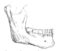
Fig. 90.—Inferior Maxillary Bone.
1, Condyle ; 2, sigmoid notch ; 3, coronoid process.
Continue to:


My Books

