Anatomy. Part 2
Description
This section is from the book "Intra-Pelvic Technic OR Manipulative Surgery of the Pelvic Organs", by Percy H. Woodall, M. D., D.O.. Also available from Amazon: Intra-Pelvic Technic OR Manipulative Surgery of the Pelvic Organs.
Anatomy. Part 2
The muscle fibers in the broad ligament are of the unstriped variety, a continuation of the outer layer of the uterine muscle, and form a flat layer between the uterus, ovaries and tubes.
The round ligaments are composed of unstriped muscle fibers from the uterus, arising from its superior angles. They pass forward, upward and outward, between the layers of the broad ligament, in front of and below the uterine tube. They enter the internal abdominal ring, pass through the inguinal canal and are inserted into the subcutaneous tissues of the labia majoria. They are supplied by the genital branch of the genito-crural nerve and are capable of electric stimulation. They are pierced through their center by a branch of the deep epigastric artery. They are from four to five inches long and contracting together tend to pull the fundus of the uterus forward and perhaps prevent retroversion in coughing, lifting or straining.
The ovaries are two flattened, elongated, oval shaped bodies about one and one-half inches long, three-fourths inches wide and about one-third inch thick. They are placed with their long axes almost vertical. Their lower extremities are attached to the uterus by the ovarian ligament, their upper extremities to the fimbriated end of the uterine tube. By their anterior margins, which look forward and outward, they are attached to the broad ligament. The free margin looks inward and backward toward the rectum. The ovaries are peculiar in that they are in reality within the peritoneal cavity. The peritoneum forming the broad ligament ceases at the attached edge of the ovary. Each ovary lies in a shallow depression on the lateral wall of the pelvis called the ovarian fossa. This fossa is bounded in front by the hypogastric artery, behind by the ureter and above by the external ilias vessels. It is slightly to the side of and in front of the rectum. The position of the ovary is variable. It perhaps never exactly regains the position from which it is displaced by the first pregnancy. It is affected by posture, the degree of distension of the bladder or rectum and by the position of the uterus. Its tendency, unless restrained by adhesions is to backward and downward displacement into the recto-uterine excavation. In its varied positions the uterine tube forms a loop around it, the inner half of the tube ascending obliquely over it, the outer half with the dilated extremity, descending and bulging out behind it, from which the fimbriae pass to grasp it.
Within the pelvis the bladder being empty, a line connecting the two ovaries would pass posterior to the body of the uterus. On the surface of the body the ovaries lie about one and one-half inches below a point about two inches toward the median line from the anterior superior spinous processes of the ilium. (Fig. 3.)
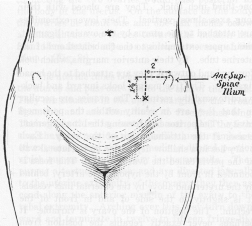
Fig. 3. Surface Location of Ovaries.
The uterine tubes begin at the superior angles of the uterus and pass outward to the side of the pelvis. They are situated in the upper margin of the broad ligament, and are from three to five inches long. Their inner third is constricted and called the isthmus, the outer portion, the infundibulum or fimbriated extremity, and the intermediate dilated portion which curves over the ovary is the ampulla. They pursue a tortuous course, and are directed laterally to the uterine extremity of the ovary; they then ascend along the anterior margin of the ovary to the tubal extremity. Arching over this they turn downward and terminate in twelve to fifteen fringe-likc processes, the fimbriated extremity that partially surrounds the ovary, embracing its free border and inner surface.
The vagina is a musculo-membranous canal leading from the vulva to the uterus. Its anterior wall which is in relation with the urethra and bladder is about two and one-half inches long. Its pos terior wall about three and one-half inches long is in relation with the perineal body at its lower fourth. Its middle two-fourths are in close relationship with the anterior rectal wall, being separated from it by a small amount of loose connective tissue with blood vessels and lymphatics. Its upper fourth is in contact with the recto-uterine excavation, and is separated from the peritoneum by a thin layer of tissue.
The walls of the vagina are in contact, a transverse section showing the canal to have the general shape of the letter H with a long transverse bar. Its axis forms nearly a right angle with the axis of the uterus when that organ is in normal position. The posterior wall extends higher up on the cervix of the uterus than does the anterior. The junction of the anterior and posterior walls of the vagina with the cervix forms the anterior and posterior fornices, respectively.
The mucous membrane of the vagina more nearly resembles skin than ordinary mucous membrane, being a stratified pavement epithelium resting upon dermal papillae.
The bladder is in relation posteriorly with the upper part of the vagina and the uterus. It is rather closely connected with the upper portion of the vagina and the front of the cervix by areolar tissue.
Its degree of fullness affects to some degree the position of the uterus.
The urethra is embedded in the anterior vaginal wall, with which its general direction coincides. It is about one and one-half to one and three-fourths inches long and about one-fourth inch in diameter. It is capable of dilation to a diameter of almost an inch.
The ureters enter the pelvis about an inch and a quarter to an inch and a half from the median line. They follow the curve of the pelvis, first downward, backward and outward; they then pass forward and toward the median line about three-fourths of an inch, lateral to the cervix of the uterus. Coursing through the upper part of the vaginal walls they reach the fundus of the bladder. They pass obliquely through the walls of the contracted bladder for a distance slightly less than one inch. They are about one inch apart, this distance increasing with distension of the bladder.
Continue to:
My Books

