Explanation Of The Plates. Plates I And II
Description
This section is from the book "A Manual Of Pathological Anatomy", by Carl Rokitansky, William Edward Swaine. Also available from Amazon: A Manual of Pathological Anatomy.
Explanation Of The Plates. Plates I And II
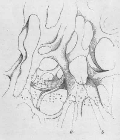
Figs. 1 and 4 represent proliferous cyst-formations from the cortical substance of the kidney, as a sequel to Bright's disease. The two figures, 1 and 4, illustrate well Roki-tansky's history of proliferous cyst-development, and at the same time what he understands by the often-occurring expression, "alveolar type or arrangement".
In fig. 1 we have the cyst in all its phases, a is a simple cyst, arising out of the expansion of the elementary granule, first into the nucleus, from this into the cell, and progressively into the cyst. But it has remained barren, and contains only a diaphanous, viscid serum within a simple cyst-membrane, b represents a parent-cyst, the early history of which accords with that of the barren cyst; within it, however, new granules have formed, and gradually become developed into vesicles or cysts containing other nuclei, until the parent-cyst has become replete with them, and from being spherical, they are rendered polyhedrical by mutual compression. In an adjoining parent-cyst, many of the filial cysts have remained barren, others contain nuclei in the act of splitting, c, c, c, c, represent another form of development of the parent-cyst. Here, again, the parent-cyst has gone through the same phases, from the elementary granule upwards. But, as the cell dilates into the cyst, a granule forms centrally to the latter and expands into a filial cyst, centrally to which a third granule opens out in the same manner; and so on.
These intra-cystic cysts in their dilatation ultimately close upon the parent-cyst, forming secondary, tertiary, and ulterior layers, to which an external, fibrous layer is generally added out of the surrounding blastema. Or this fibrous coat accrues in the alveolar shape. Fig. 1 affords several examples of this. It is, however, better seen in Fig. 4. - a is the fibrous sheath in progress of development out of d, the elongated and caudate nuclei coursing around the parent-cyst or aggregation of parent-cysts. They eventually break up into the requisite fibres, e is to represent the point-molecule, within an amorphous blastema, out of which the nuclei (6) form. They are at first spherical, afterwards elongated, and ultimately broken into fibrillation. This constitutes what the author designates as the "alveolar type or arrangement." It is, however, still better defined in, Fig. 2. which represents cyst-formation in a medullary carcinoma. From the carcinomatous framework a bulb-like excrescence is thrown out, within the extremity of which a parent-cyst forms and becomes replete with filial cysts, each containing a central nucleus. This parent-cyst surrounds itself with a broad marginal area of blastema, within which elongated, caudate nuclei course round the cyst in several tolerably regular circles or series - the rudiments of a dense fibrous envelope. Such is the "alveolar type," which applies to the fibrous fabric of follicle walls as well as to those of cyst-formations. (See "Cyst and Alveolus.")
Fig. 1.
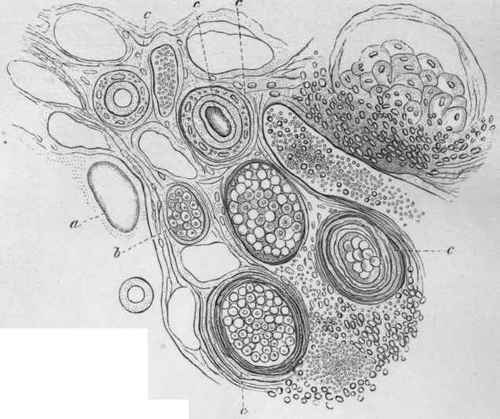
Fig. 2.
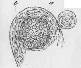
Fig. 4.
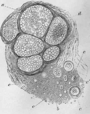
Fig. 3 represents a transverse section of a colloid cancer, a is an older portion of densely fibrillated fibro-membranous structure. c is a transverse section of a more recent fibro-membranous stroma; b, a transverse section of the colloid warp which intertwines with the said fibro-membranous stroma. (See p. 220).
Fig. 3.
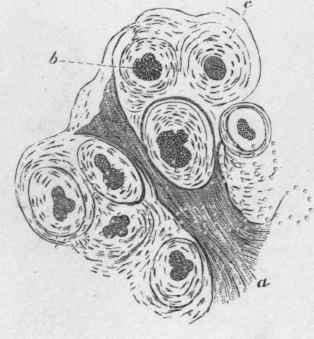
Fig. 8 represents the multilocular, fibro-membranous stroma of colloid cancer deprived of its colloid contents. (See p. 221).
Fig. 8.
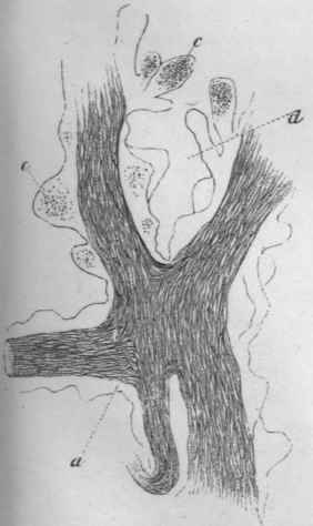
Figs. 5,6, and 7, represent so many stages of the development of medullary carcinoma. They are severally described in the same order in which they are here numbered, at pp. 220 and 221".
Fig. 5.
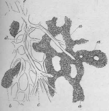
Fig. 6.
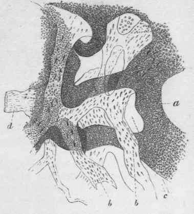
Fig. 7.
Figs. 1, 2, and 4 are magnified by 90 diameters, the five remaining figures by 400 diameters.
Several of the figures here given are embodied from Rokitansky's "Essays," in Mr. Paget's admirable "Lectures on Surgical Pathology," vol. ii.
Figs. 1, 2, and 4, are derived from Rokitansky's Essay on "Cyst and Alveolus," read before the Imperial Academy of Sciences, at Vienna, in 1849; figs. 3 and 8 from his Essay on "Colloid Cancer," published in 1852; figs. 5, 6, and 7, from a thesis of his on "Cancer-stromata," also published in 1852.
Continue to:
My Books

