


2. Ascaris Lumbricoides
Description
This section is from the book "A Manual Of Pathology", by Joseph Coats, Lewis K. Sutherland. Also available from Amazon: A Manual Of Pathology.
2. Ascaris Lumbricoides
The common round-worm is probably the commonest entozoon in the human subject. It occurs very frequently in children, and inhabits chiefly the small intestine. In its colour and general appearance it resembles the common earth-worm (see Fig. 194). It measures 6 to 16 inches in length, is marked by transverse striae, and tapers to both ends. Like other nematodes it possesses an intestinal canal from end to end. The female produces a large number of oval eggs which have a dense shell (see Fig. 195). The ova are discharged from the intestine and may be found in the faeces.

Fig. 194. - A clump of ascarides, large and small, from the intestine.
The worm mostly occurs singly or in pairs, but is frequently present in considerable numbers up to one or two hundred. From the intestine it may pass into the stomach and be vomited, or may be discharged per anum. It has been known also to pass up the oesophagus and into the-nostrils and sinuses of the head, or by the larynx into the bronchial tubes. Sometimes it penetrates into the bile ducts, which it may obstruct, or passes through the intestinal wall into the peritoneal cavity. In cases where they have perforated into the peritoneum they have given rise usually to local abscesses pointing chiefly near the umbilicus or groin. More rarely they have led to general peritonitis.
In the intestine the irritation of the worms produces catarrh, and by reflex action this is supposed to lead to certain nervous symptoms. When present in large numbers, the worms are sometimes rolled up in a ball, and in this condition they may obstruct the intestine.
Ascaris Mystax
This is a small round-worm which occurs in the cat, and is said to be always present in the intestine of that animal.
Oxyuris Vermicularis Or Thread-Worm
This is an exceedingly common parasite. It is white in colour, and the male measures about an eighth of an inch, and the female about three-eighths in length. It possesses an alimentary canal from end to end. The eggs are oval, and have a dense shell. The animal inhabits mostly the large intestine. It is stated by Zenker aud Heller that the mature female is in the large intestine, the males and young being in the small. The worm often wanders, especially during the night, to the neighbourhood of the anus, where it produces itching. Sometimes it passes over to the vagina, and up into it. It produces catarrh of the bowel, and, as in the case of the ascaris, nervous symptoms are ascribed to it.
Trichocephalus Dispar (The Whip-Shaped Worm)
It is of frequent occurrence in the caecum and neighbouring parts of the intestine. It measures 1 1/2 to 2 inches in length and has the peculiarity that the anterior portion is much thinner than the posterior, forming a long thread, like the lash of a whip (see Fig. 196), which is buried in the mucous membrane. It seems to produce no special symptoms. The eggs possess a brown shell. The embryoes have been traced in water and moist earth.
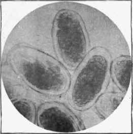
Fig. 195. - Ova of ascaris lumbricoides. x 280.
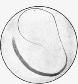
Fig. 196. - Trichocephalus dispar or whip-shaped worm from the ceecum. x 3.
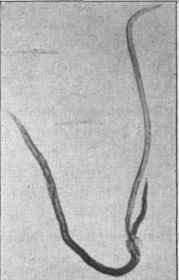
Fig. 197. - Doehmius duodenalis, male and female, x 3.
Dochmius Duodenalis (Strongylus Duodenalis. Anchijlodomum Duo-Denale)
This worm is rarely met with in this country, but occurs in Egypt, Italy, and tropical lands. It has been found frequently among the workers at the St. Gothard Tunnel in Switzerland. It is .a third to half an inch in length (Fig. 197), and it possesses a mouth armed with four strong teeth. By means of these it fixes itself on the mucous membrane where it sucks the blood. When present in considerable numbers, as it often is among the valvulae conniventes, it may give rise to considerable loss of blood and serious anaemia (Anaemia Egyptorum).
Strongylus Gigas
This is a large worm, reaching a length of over a yard, and a thickness of about three-eighths of a inch. It has been met with a few times in the pelvis of the kidney in man, and more frequently in the kidney, bladder, lungs, and liver of dogs.
Round-worms belonging to the family of the Strongylidse are frequent parasites in the lower animals. These inhabit various parts of the body, but there are two situations in which they are of special frequency, namely the lungs and the arteries.
Pulmonary and bronchial Strongylosis is the name applied to the conditions produced by various strongyli when resident in the bronchial tubes and lung-parenchyma. No less than eight species of the genus strongylus have been distinguished (Neumann), and the animals affected include the sheep, goat, deer, ox, horse, ass, hare, rabbit, and cat. The disease is seen in its most characteristic form in the sheep. There are two species met with in this animal, namely, the Strongylus filaria which inhabits the bronchi and gives rise to bronchitis and the Strongylus rufescens which penetrates to the lung alveoli. In the latter case a pneumonia is produced, each worm being the centre of a local inflammation. There are thus nodules which have a superficial resemblance to tubercles.
Aneurysms are frequent in the horse and ass in consequence of the action of one of the Strongylidre, the Scleroatoma armatum or equinum. The worm is first present in the large intestine, from which it penetrates to the arteries which come off from the abdominal aorta, chiefly the cceliac and mesenteric arteries, very rarely the aorta itself. The worm produces thrombosis and aneurysmal dilatation of the vessels. From the discharge of portions of thrombi, embolism in the arteries of the intestine results, giving rise to colic, which is a frequent symptom in the horse. The aneurysm's are present in about 90 per cent, of horses, and are equally frequent in asses.
Continue to:


My Books

