


2. Colloid Ovarian Cystoma
Description
This section is from the book "A Manual Of Pathology", by Joseph Coats, Lewis K. Sutherland. Also available from Amazon: A Manual Of Pathology.
2. Colloid Ovarian Cystoma
This form is of much more frequent occurrence and vastly more important. To this class of cysts the name Cystoma is properly applied, because they arise by a distinct new-formation, there being first produced a preparatory tissue, which goes on to the formation of the cysts. The preparatory tissue is glandular in character, and hence the names Adenoma and Adenocystoma are sometimes applied. This group includes the characteristic Multilocular cysts of the ovary.

Fig. 460. - From a colloid ovarian cystoma. Gland-like tissue and the beginning of cysts, x 70.

Fig. 461. - Portion of wall of colloid ovarian cyst. The lining epithelium is mostly in the form of goblet cells, x 360.
These cysts form bulky tumours, and while the tumour itself is formed of a number of larger and smaller cysts, there is nearly always, in the walls of these, more or less solid material which shows various stages in the process of cystic formation. The tumour, sometimes of enormous size, represents the Ovary as a whole, and is not merely something added to it, the external covering of the cyst corresponding strictly with the surface of the ovary.
The solid tissue in the wall of the cysts shows the various stages in the process of development. It shows, microscopically, a glandular structure (see Fig. 460) in the form of tubular canals lined with cylindrical epithelium. These gland-like structures often project into cysts, and altogether they show a very striking power of new-formation. The transformation of the gland-like tissue into cysts is readily seen in many cases. The cylindrical epithelium presents very markedly the goblet form seen in mucous glands (Fig. 461), and it secretes a mucous or colloid material, which accumulates in the cavity and distends it more and more into a globular cyst. We have therefore, in the walls of the cysts, very commonly large numbers of smaller cysts which take their shape by mutual pressure, as in Fig. 462. Cysts formed in this way project into the already existing cysts as they grow, and may afterwards burst and become flattened out on the wall of the older cyst. As all the cysts have had a similar origin, they are lined with epithelium, whose function it is to secrete a colloid or mucoid fluid, and so the cysts, once formed, have an almost continuous tendency to increase.

Fig. 462. - Section of portion of a projection on internal surface of ovarian cyst. It consists of a congeries of variously sized smaller cysts, x 20.
As regards the Origin and Significance of the glandular tissue, authors are, generally agreed that, as we have here essentially to do with glandular epithelium, its origin is to be referred to the primordial epithelial structures of the ovary. The primordial ovary consists of a layer of epithelium on the surface of a connective-tissue projection. The ova form by the penetration inwards or reduplication of this superficial epithelium, which at first forms a series of communicating channels or follicles which Waldeyer compares to a cavernous tissue. By the constriction of these canals the ova are formed, it being doubtful whether the cells of the membrana granulosa which lines the Graafian vesicle are derived from this epithelium or from the connective-tissue stroma. The glandular formation in ovarian cystoma may be regarded as a pathological and exaggerated recurrence of the fcetal condition.
The Naked-eye appearances of the colloid cystoma are generally quite characteristic, and;may be inferred from Fig. 463, in which a tumour is shown in section after hardening. The tumour represents an ovary, and its outer covering is the outer covering of the ovary with its layer of endothelium. The surface is usually smooth and very often free from adhesion to neighbouring structures. In shape the tumour is generally more or less globular, but not infrequently it is lobulated on the surface and evidently composed of several cysts. Although the cyst is for the most part obviously multilocular, yet one of the cysts sometimes attains such a preponderating size that the tumour is apparently unilocular. In these cases examination will show the existence of other cysts flattened out in the wall, or collected here and there in clusters. Towards the base of the tumour, but also in many cases at various places, there are solid or semi-solid masses in which cysts are in process of development in the way already described. The size of the tumour is frequently very great. On cutting into it there escapes from the cysts a sticky brownish or yellowish fluid, which is tolerably clear unless some of the secondary changes to be presently described have occurred in it. If, as is usually the case, there are many cysts, they take shape by mutual pressure against each other, while the general globular outline of the tumour as a whole is preserved.
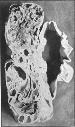
Fig. 463. - Colloid ovariuu cystoma in section after hardening. There is one large cyst to the right which has become shrunk. The rest of the tumour consists of cysts of various sices, with some solid tissue. (About one-third the natural size).
As the ovary hangs free in the peritoneum the tumour also generally expands outwards freely, and preserves a comparatively narrow attachment, called the Pedicle. In ovarian cysts as excised the ala vesperti-lionis will generally be found attached and nearly normal, and the parovarium is often unusually well preserved. The expansion of the tumour is very rarely upwards from the ovary into the ala vesperti-lionis. If it be so, then the tumour is more sessile and the Fallopian tube may be stretched over its summit.
Secondary changes occur which concern chiefly the contents of the cysts. It seems almost a normal condition in large cysts that the internal wall should present collections of cells in a state of Fatty degeneration, these cells being often in many layers. Cells also pass into the fluid, so that in all ovarian fluids cells are to be found of round shape and with oil drops in them. Many of these cells are doubtless leucocytes which have emigrated into the cysts. The number of these fatty cells may be greatly increased, and, as many of them in that case break down, we may have a fluid which is turbid from the presence of numerous fatty cells and free oil. ' The fluid is also pale like a fatty emulsion, and in many cases it resembles pus in its naked-eye appearance. This change is most likely to occur in old and large cysts, but it is not infrequent in younger and smaller ones.
The free fat, if long retained, forms Crystals of cholestearine, and in fact these crystals are frequently found in the fluid of cysts which otherwise are not strikingly altered.
Haemorrhage occasionally occurs into the cysts, and this will cause the fluid to be turbid and deep brown or red in colour. There may also be masses of softened fibrine in the cavity. Sometimes, however, the fluid has a dark brown colour without haemorrhage.
Inflammation of the cyst-wall is not of very frequent occurrence. There may be an acute suppurative inflammation, so that the contents become mixed with pus and assume more and more of the purulent character. With this there is generally an acute inflammation of the surface, with fibrinous exudation and adhesion to neighbouring structures. If the suppuration continues there is apt to be perforation of the pus into the abdominal cavity with resulting fatal peritonitis. A chronic inflammation is more common, causing adhesion of the cyst to neighbouring parts, and these adhesions may be very extensive and firm.
Perforation or Rupture of the individual cysts which compose the tumour is not infrequent. It has been already stated that coalescence of cysts by rupture of their adjacent walls is a regular process in the course of growth. A rupture externally is much less frequent. It occurs by mechanical violence, by necrosis of parts of the wall from interference with the circulation, from inflammation, or from penetration of the wall by the growth of the secondary cysts.
The rupture occurs usually into the peritoneum, but it may be into the rectum or bladder, inflammatory adhesions having previously formed. According to Bland Sutton a cystoma may rupture and continue to secrete colloid matter which passes into the peritoneum, and may collect there in large quantity. The colloid matter in the peritoneal cavity sets up inflammation which may be somewhat acute. If it lasts for some time there is great .thickening, especially of the omentum, which contains colloid masses, and looks like a mass of boiled sago (Doran and Olshausen).
Continue to:


My Books

