


2. Myoma
Description
This section is from the book "A Manual Of Pathology", by Joseph Coats, Lewis K. Sutherland. Also available from Amazon: A Manual Of Pathology.
2. Myoma
The myoma is very infrequent in the vagina and in the cervix of the uterus, but is extremely frequent in the body of the uterus. It occurs in from 10 to 20 per cent, of women beyond 20 years of age, and in about 40 per cent, of those above 50.
The myoma presents great varieties in size, in number, and in the details of its structure. There may be a single small tumour about the size of a pea, or a large growth weighing more than fifty pounds. There are frequently several tumours present, and there may be fifty attached to the same uterus. The tumour is usually hard and on section fibrous in appearance.
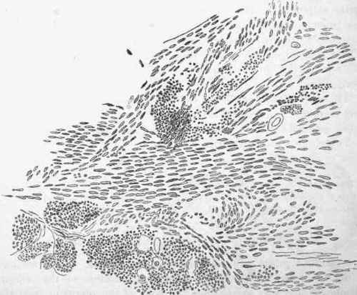
Fig. 402. - Section of a myoma of the uterus stained with carmine. The muscular nuclei are seen in longitudinal and transverse section, x 350.
In structure the chief variations are in regard to the proportion of muscle, connective tissue, and vessels. The muscle is in bundles which to the naked eye often give a concentric arrangement to the cut surface (see Fig. 454). There is under the microscope, the usual arrangement of the nuclei in the muscular bundles, as shown in Fig. 452. The character of these nuclei and their arrangement are sufficiently distinctive of the tumour.
Connective tissue is present between the muscular bundles; sometimes it is dense and, by rendering the tumour compact, gives it a very hard fibrous character. In some cases the connective tissue increases out of proportion to the muscle and a process of induration akin to cirrhosis -occurs.
The vessels are usually rather sparse in the myoma, but sometimes great dilatation of the blood-vessels occurs, so as to give a cavernous character to parts of the tumour, a condition indicated by the name Myoma telangiectodes. In other cases there is a dilatation of the lymphatic vessels, leading to a condition called Myoma lymphangi-ectodes.
Secondary changes are very liable to occur in the myomas, especially as the tumours frequently grow to large dimensions and are liable to changes in position which interfere with the blood-vessels.
Cysts not infrequently form in them. These are sometimes from dilated lymphatics, but more frequently from softening of the tumour tissue. Large cysts are thus formed in the midst of large myomas, and may even give rise to a feeling of fluctuation.
(Edema and Haemorrhage are not uncommon, the latter especially, and more particularly in the tumours with dilated blood-vessels.
Calcareous infiltration occurs in two different forms. A part of a large tumour may, from obstruction of vessels, be cut off from its blood supply and become obsolete, or a tumour may be separated as a whole and become calcified. In the latter case lime salts may infiltrate it and they are deposited in all the constituents, the muscle-cells, walls of the blood-vessels, and connective tissue (see Fig. 53, p. 154, which is from the calcified part of a myoma).
Separation and Transplantation are not uncommon in the myoma. The subserous myoma, after becoming pedunculated, may acquire vascular connections with the omentum or other part of the peritoneum and ultimately become detached from the uterus. In a case observed by the author a tumour seven inches in length was attached to a very long great omentum and was movable in the abdomen.
The submucous forms also sometimes become detached, and may be found in the cavity of the uterus; they may either be discharged or retained.
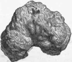
Fig. 453. - Calcified myoma of uterua found lying in the cavity. Half the natural size.
The detached or transplanted myoma is apt to undergo calcification.
In a specimen sent to the author by Dr. Chapman, of Hereford, and depicted in Fig. 453, a myoma was found lying loose in the uterus. It had externally a firm ?hell which had to be sawn through in order to divide the tumour. The calcification extended to intersecting trabecular, which divided the tissue into loculi. In these loculi soft tissue existed which had the microscopic characters of that of the ordinary myoma. The specimen is preserved in the Museum of the Western Infirmary.
The myoma originates in the muscular substance of the uterus, and it may remain in the wall or become displaced outwards or inwards. Hence there are three varieties.
(a) The Subserous myoma, originating in the external layers of the uterus, passes outwards as it grows, and pushes the peritoneal coat before it. In this way it frequently becomes pedunculated. The subserous form is often multiple, and as, from its situation, it is protected, the tumour may grow for many years undisturbed, and reach very large dimensions. Such large myomas may be mistaken for ovarian tumours and excised as such, and this is the more likely as cysts not infrequently occur in them.
(b) The Interstitial or Intra-parietal myoma in its growth involves the wall of the uterus, and may cause enormous enlargement of the organ (see Fig. 454; the actual tumour measured 9 1/2 inches in long diameter). This form develops mostly at the fundus, and usually occupies the posterior wall. The tumour and greatly enlarged uterus may form together a very bulky mass, which as a whole is liable to be mistaken for ail ovarian or other tumour. The author has met with several cases in which the tumour and uterus were excised under this belief. In one of these the tumours seemed to be multiple and the uterine wall could not be distinguished from tumours, the greatly enlarged cavity of the uterus being surrounded by irregularly lobulated masses of.muscular tissue. In this case it looked as if the uterus as a whole had undergone an irregular hypertrophy, or had grown into a massive tumour.

Fig. 454. - Giganticintraparietal myomaof uterus. It occupied the posterior wall and somewhat distended the os uteri, whose lips were almost of papery thickness. It partly projected into the vagina (a). The greatly dilated cavity of uterus (6) lay in front, c, urinary bladder; d, rectum.
(c) The Submucous myoma is the form which most frequently comes under the notice of the practitioner. Arising in the wall of the uterus, it passes inwards, pushing the mucous membrane before it, and from the action of gravity it tends to become pendulous. The submucous myoma, therefore, very often presents itself as a polypus (the so-called Fibroid polypus), and it may have a very narrow neck. It very often arises at the fundus, and may grow to such dimensions as to fill the uterus, and hang down through the cervix into the vagina (see Fig. 455). The mucous membrane covering the tumour is subject to irritation, and there is frequently Hemorrhage and Ulceration, even with sloughing in some cases.
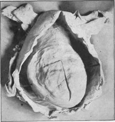
Fig. 455. - Largo submucous myoma attached near the fundus and distending uterus and vagina.
Continue to:


My Books

