


6. Retrograde Changes In Arteries
Description
This section is from the book "A Manual Of Pathology", by Joseph Coats, Lewis K. Sutherland. Also available from Amazon: A Manual Of Pathology.
6. Retrograde Changes In Arteries
We have already referred to retrograde changes in connection with atheroma. Some more independent forms have still to be considered.
(A) Fatty Degeneration Of The Intima Of Arteries
This condition is apt to be mistaken for atheroma. We frequently see, especially in the aorta, yellow markings slightly raised above the internal surface, and having the appearance of superficial figurings. They are to be found in the aorta, pulmonary artery, and other parts of the arterial system. If a portion of such a patch be examined under the microscope by removing a thin layer by a section parallel to the surface, it will be found that the condition consists in a fatty degeneration of the cells of the intima. The flat branched cells are demarcated by the presence in them of abundant fat drops. If the fatty degeneration is much advanced then the intercellular substance becomes also the seat of fat drops, and the cells are no longer demarcated. It sometimes happens that when the degeneration is very advanced the little patch softens and an erosion forms. This erosion is very superficial, and is not to be mistaken for the atheromatous ulcer.
This condition is sometimes met with in the bodies of comparatively healthy persons; we have already seen that in anaemic and emaciated persons it is frequent, and it is to be classed in the same category as fatty degeneration of the muscular tissue of the heart.
(B) Calcareous Infiltration Of Arteries
We have already seen that in atheroma there is very frequently a calcareous infiltration of the affected structures, and it has been stated that individual peculiarities appear to play, an importarrt part here. In some cases a more independent calcareous depositiorr occurs, and here irrdividual peculiarities are of still greater consequence. Calcification of the middle coat is the most frequent and important form. This is very frequently associated with atheroma, but it is noteworthy that the atheroma and calcareous deposition very often affect different arteries or different parts of the same artery. We may have, for instance, atheroma of the aorta and calcification of the middle coat in the femoral and smaller vessels; or there may be atheroma as well as calcification in the femoral, but in their extreme degrees the two are present at separate parts of the artery. For instance, Fig. 241, p. 482, and Fig. 249 are from the same femoral artery, and from parts near each other; but the one shows atheroma with fatty degeneration, while the other exhibits very advanced' calcareous infiltration of the middle coat. It may here be remarked that calcification affects by preference the arteries of medium and smaller size. It is often very pronounced in the femoral and brachial, and extends to those of smaller size, but not to the finest arteries.
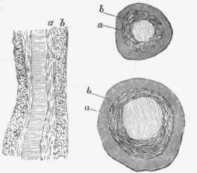
Fig. 247. - Endarteritis obliterans in arteries of kidney: a, thickened and fibrous internal coat; b, middle coat.
The deposition occurs in most cases primarily into the muscular fibre cells of the middle coat, and at first marks these out by the presence of fine opaque granules (see Fig. 248), but the author has met with a case in which the elastic lamina of Henle was the primary seat of deposition. The granules flow together till a patch is formed of an opaque appearance, but confined to the middle coat. As the calcareous deposition increases, the patch sometimes assumes a crystalline appearance, and the material may become broken and irregular, as in Fig. 249. This condition necessarily gives greatly increased rigidity to the walls of the arteries, and the feeling of rigidity so often felt in the radial and other smaller arteries is mostly due to this cause, and not to atheroma. The fact, however, that calcification of the middle coat is so often associated with atheroma renders this rigidity to some extent an indication of the existence of atheroma in the larger arteries. # To the naked eye the calcified parts frequently manifest themselves by the appearance of a circular opaque striation, visible especially when the artery is laid open and viewed from within. The appearance is better seen if the artery be allowed to dry partially, as then the normal tissue becomes more transparent and the chalky structures more prominent. In that case complete or partial rings are seen, not unlike the irregular cartilaginous rings of the bronchi, but smaller. Another method of rendering the calcification prominent is to tear off the internal coat, which is usually somewhat loosely attached.

Fig. 248. - Calcareous infiltration of the middle coat (6) in an artery, early stage, with atheroma (a). At the edge of the affected part, the general outline of the muscle fibre cells can be made out. x 60.
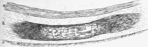
Fig. 249. - Calcareous infiltration of the middle coat of an artery. The lime salts have aggregated together so as to produce a crystalline appearance. This was taken from the same artery as Fig. 241. x 22.
The calcified middle coat sometimes undergoes a true Ossification, so that spicula of bone may be mixed with calcareous masses. The ossification is secondary to the calcareous infiltration, and is always small in extent compared with the latter. It is really the result of a chronic inflammation around the calcareous masses, the new-formed connective tissue, penetrating amongst these, becoming transformed into true bone by reason of the rigidity of the structures and the abundance of lime salts.
The author has observed a similar development of true bone in a collapsed hydatid cyst in the liver. In this case the new-formed connective tissue had penetrated amongst the degenerated products of the parasite, and in some parts had developed osseous tissue with regular lacunae and canaliculi.
The calcareous infiltration of the middle coat renders the artery peculiarly rigid, and, when extensive, induces hypertrophy of the left ventricle just as rigidity of the aorta does. The rigid vessels are also liable to dilatation, as in the case of the aorta. But besides that, the calcified middle coat is brittle, and affords a much less effective resistance to the distensile wave of blood. An additional strain on the circulation, or some special movement of the body, may break the brittle coat and directly lead to aneurysmal dilatation of the artery. It is not unlikely that peripheral aneurysms are frequently induced in this way, and especially those of the popliteal region, where the artery is peculiarly liable to mechanical injury from the movements of the limb.
A peculiar Calcification of arteries by metastasis has been described. We have already seen that when great destruction of bone is occurring the lime salts may be deposited in distant parts, the lungs and mucous membrane of the alimentary canal being the chief seats. Kuttner has described a case in which the incrustation occurred in the arteries. The deposition increased in amount the further removed the arteries were from the heart, and it was especially manifest where the current was permanently impeded, as where a small branch issued from a comparatively large stem. In this case it was the intima that was incrusted, and it was peculiar that the veins were not in the least affected. Apart from the incrustation, the arterial coats were apparently normal. The source of the lime salts was an acute caries of the vertebral column from the first dorsal to the last lumbar. In relation to the pathology of the case, it is to be added that there was a purulent interstitial nephritis, so that the excretion of lime salts was presumably impeded. It is remarkable that in this case the metastasis was to the arteries, and that the lungs and the mucous membrane of the intestine were entirely free.
(C) Amyloid Disease
This is frequent in the walls of arteries in various organs. (See page 146, and under Kidneys, Spleen, Liver).
Continue to:


My Books

