


Atheroma. Chronic Endarteritis. (Arteriosclerosis, Endarteritis Deformans, Endarteritis Nodosa). Continued
Description
This section is from the book "A Manual Of Pathology", by Joseph Coats, Lewis K. Sutherland. Also available from Amazon: A Manual Of Pathology.
Atheroma. Chronic Endarteritis. (Arteriosclerosis, Endarteritis Deformans, Endarteritis Nodosa). Continued
Calcareous infiltration is a frequent result in the atheromatous patch. The dense tissue of the patch may become infiltrated with lime salts, with or without a preceding fatty degeneration. This is peculiarly the ease when the tissue is very hard and dry*. We shall see afterwards that some individuals present a very great tendency to the deposition of lime salts in the walls of their arteries, and there are cases of atheroma in which this tendency is very marked, so that with comparatively little atheromatous thickening there may be very extensive calcareous deposition. At first the salts are deposited in fine granules, and in the deeper layers of the patch, but as time goes on these aggregate into consistent masses, so that we have Calcareous plates of various forms and sizes, sometimes attaining to a square inch in area. These plates, having abrupt edges, not infrequently tear through the remaining layers of the intima, and present an edge or angle inside the vessel. They may even become to a considerable extent separated, and hang into the calibre attached only by a strip of intima like a hinge. The rough calcareous edge very commonly induces a deposition of hi>iinc, and the thrombus thus formed may subsequently get detached, and form an embolus. The determination of the preponde ranee of the fetty or calcareous change appears to depend on individual peculiarities. Both forms are very frequent in the aorta. The fatty change preponderates greatly in the cerebral arteries. The aorta is sometimes greatly altered by the atheroma and calcareous deposition, as shown in Fig. 243.
Besides these, which may be regarded as the primary lesions of atheroma, there are frequently present certain further changes which liivc Keen variously interpreted as parts of the primary lesion or as Seconday results. . These concern chiefly the media and the adventitia. There is no doubt that in atheroma of the aorta there are very commonly inflammatory manifestations in the two outer coats, and some authors have regarded this as an indication that the atheroma is merely part of a general inflammation of the vessel-wall. But these manifestations are usually absent in atheroma of the smaller vessels, and may be awanting in the aorta itself. They are either the result of the atheroma, or else they are conditioned by those changes in the circulation (increased pressure) which we have seen to take part in the causation of atheroma.
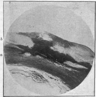
Fig. 242. - Atheroma of aorta: a. middle coat; b, atheromatous internal coat. There is a marked opacity of the intima in its deeper parts from fatty degeneration, and it impinges on the media. The fatty structure has come to the surface and caused an atheromatous ulcer, x 8.
The atheromatous patch, forming a hard nodule, impinges on the media, and not infrequently seriously interferes with it. There may be a simple thinning of the media from within, along with fatty changes which may manifest themselves in the muscle cells of this coat (see Fig. 244). There may be, on the other hand, traumatic lesions, sometimes resulting in rupture of the elastic lamina of Henle. This injury and rupture brings about a process of repair, so that we have the usual formation of vascular" inflammatory tissue in the media going on to cicatrization. Moreover, the atheromatous patch is largely in the condition of a piece of dead or obsolete tissue, and, on the principles already expounded, it is liable to be eaten into and replaced by vascular tissue. The deeper parts of the patch are frequently thus vascularized, especially • 71 the media has been injured. These are true inflammatory changes in the intima, and are different from the atheromatous process. They are often continuous with the similar changes in the media and adventitia. These lesions are of importance in relation to the causation of aneurysms (see further on).

Fig. 243. - Portion of thoracic aorta, almost continuously atheromatous, and with many calcareous plates.
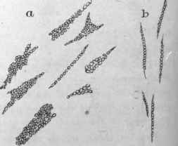
Fig. 244. - Fatty degeneration in atheroma: a, from internal coat; 6, muscle cells from middle coat, x 350.
(C) Effects Of Atheroma On The Circulation
There are four alterations which it produces, each of which may, according to circumstances, have important effects on the circulation. The disease causes narrowing of the calibre, interference with the muscular contractility, rigidity, and sometimes actual injury to the wall.
The Narrowing of the vessel will be of little consequence in such large arteries as the aorta, but in the case of the cerebral vessels, the cardiac arteries, and those of the legs, the interference with the circulation may be considerable (see Fig. 245). We have seen this illustrated in 'the case of the coronary arteries of the heart, especially when thrombosis supervenes, where the disturbance of the circulation sometimes produces severe angina, and may lead to sudden death, and it is no less potent as a cause of disease in the cerebral arteries.
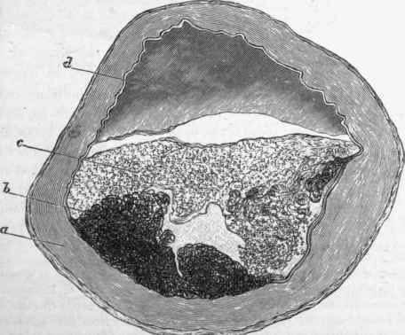
Fig. 245. - Atheroma of cerebral artery. The greatly thickened internal coat is seen. In its substance are dark masses (b) in which margarine crystals were found. On the surface is a paler layer (c) consisting of partially organized thrombus; (d) blood occupying the remaining calibre, x 34.
The Interference with the contractility will also affect mainly the arteries of smaller dimensions, and in the case of the arteries of the brain, the absence of that control of the circulation which is afforded by their varying calibre may lead to serious consequences.
Rigidity and weakening of the wall are very important consequences of atheroma, especially in the aorta, where they are important factors in the causation of aneurysm. The immediate consequence of rigidity of the aorta is that during the systole of the heart the vessel does not dilate, and at the end of the systole it does not recoil, so that the force of the elastic recoil is lost to the circulation, and in distant parts there is apt to be more or less stagnation. As a result of this we have hypertrophy of the left ventricle, which is often very marked in cases where much calcareous infiltration exists. The hypertrophied ventricle sending the blood forcibly into the rigid aorta produces commonly a diffuse dilatation of the arch. The influence of atheroma in producing aneurysm will fall to be considered subsequently.
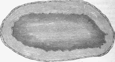
Fig. 246. - Transverse section of normal cerebral artery to contrast with Fig. 256. x 34.
The time of life at which atheroma is most frequent is a point of some importance in relation to the causation of aneurysm. According to Rokitansky, it is commonest between the ages of forty and sixty. It is still pretty frequent down to thirty years of ages, but rapidly diminishes in frequency from that age downwards. It is extremely rare under twenty years, and when it does occur it is mostly in connection with congenital anomalies of the great vessels or heart, such as stenosis of a main stem with defect of the septum, etc.
Continue to:


My Books

