


C. The Encephalon
Description
This section is from the book "A Manual Of Pathology", by Joseph Coats, Lewis K. Sutherland. Also available from Amazon: A Manual Of Pathology.
C. The Encephalon
C. The Encephalon. - Anatomical Introduction, arrangement of fibres and centres. Functions of convolutions. Arteries of brain. I. Malformations. 1. Congenital smallness, microcephalus. 2. Hypertrophy. 3. Heterotopia. II. Injuries. Laceration of brain. III. Lesions affecting the Circulation. 1. Hyperemia, anaemia, and cedema. 2. Occlusion of arteries; by embolism or thrombosis; effects, including Softening of the brain substance; occlusion in chorea. 3. Thrombosis of the cerebral sinuses. IV. Cerebral Haemorrhage. 1. From large arteries, chiefly due to aneurysm, sometimes to atheroma; influence of increase of blood-pressure. 2. From nutrient arteries, may be from miliary aneurysms or atheroma. 3. From capillaries, chiefly complicating other forms. Appearances in haemorrhage, and results; the apoplectic cicatrix and cyst. V. Inflammation. 1. Acute localized encephalitis. 2. Abscess of the brain. 3. Chronic localized encephalitis. 4. Diffuse encephalitis, (a) in fevers, (6) in dementia paralytica, (c) in hydrophobia, (d) in tetanus. VI. Atrophy and Degenerations. Senile atrophy and other forms; secondary degenerations. VII. Tuberculosis and Syphilis. 1. The tubercular tumour. 2. Syphilis of brain and membranes; disease of arteries may lead to softening. VIII. Tumours, chiefly glioma and sarcoma, and Parasites.
Anatomical Introduction
A knowledge of the course of the fibres in the encephalon is even more important than in the spinal cord and medulla, as these are liable to be interrupted in a more isolated "fashion. It is of great consequence that the pathologist should have an acquaintance with the general relations of parts so as to identify the position of lesions. In this section nothing more is attempted than to indicate these general relations, the more intricate particulars being left to the special works on anatomy. In the actual work of post-mortem examination, it is important to note the exact locality of lesions on the spot. To assist in this, tracings may be made of the figures which accompany this section, or of similar ones, the positions of the lesions being entered in shading.
In the medulla oblongata, as we have seen, motor fibres, which have been in the lateral columns of the cord, come forward and form the anterior pyramids, in which they decussate. The sensory fibres are now behind, and they remain posterior to the motor in all the succeeding parts. In the Pons varolii (Fig. 320) the motor fibres are in front, forming bundles (as shown in lower half of figure), but overlaid by the transverse fibres from the cerebellum. Besides these fibres, which are mainly in the anterior half of the pons, there are grey nuclei (shown in upper part of figure), which continue up the series in the floor of the fourth ventricle. In the pons the principal nuclei are those of the sixth and facial, the motor and sensory roots of the fifth, and one of the nuclei of the auditory nerve. It is to be remembered also that the fibres of these nerves in part at least traverse the pons, and are liable to be involved by a lesion, even when the nuclei are not reached.

Fig. 320. - Pons varolii. In lower half the transverse section of nerve bundles continued upwards from the cord is seen, the pyramidal tract being in front (or lower in figure). The Roman numerals and letters indicate the nuclei of cerebral nerves. (Quain).
In the Crura cerebri, or cerebral peduncles, the motor fibres are in front and internal, forming the greater part of the crusta. Sensory fibres occupy about the external fourth of the crusta. Behind the crusta is the locus niger, behind which again is the tegmentum, containing a rather complex mass of fibres and grey matter.
Above the crura the fibres are continued upwards, the motor still anterior and the sensory posterior, in the mass of white substance called the Internal capsule. It is here necessary to be somewhat more minute in the description of the relation of the structures, as these relations are of great importance. When one of the lateral ventricles of the brain is opened, certain masses of grey nervous tissue are seen in its floor. In front there is a long brown prominence, rounded anteriorly, and tailed behind. This is the Nucleus caudatus, which is often designated the corpus striatum, although really only one piece of it. Behind the nucleus caudatus is the Optic thalamus, which is more bulky and rounded. It is to be remembered that in opening the lateral ventricle almost no nerve fibres need to be cut except the commissural ones of the corpus callosum. The great masses of nerve fibres passing upwards are as it were pushed outwards by the lateral ventricle, and we have to cut into its floor in order to reach them.
From the accompanying figure, which represents a horizontal section of the brain just below the floor of the lateral ventricle, the relations of parts may be gathered (Fig. 321). Beneath and outside the nucleus caudatus (NC), and continuous above and externally with the great central white substance of the hemispheres, the corona radiata, there is a mass of white substance. This is the Internal capsule, in which may be distinguished an anterior division IK', a posterior division IK, and a middle part, the knee K. To the outside of and beneath the internal capsule lies a mass of grey substance, the Nucleus lenticularis (LN), which on section has a triangular shape with the base turned outwards, and is seen to be divided into three pieces (shown on right side). It is generally regarded as a part of the corpus striatum and motor in function. The nucleus lenticularis extends a considerable distance from before backwards, and in its posterior parts the internal capsule (IK) lies between it and the optic thalamus, which has now largely taken the place of the nucleus caudatus. Outside the nucleus lenticularis there is a narrow band of white substance (EK), the External capsule, the capsules being named from their relation to the nucleus lenticularis. Outside the external capsule again and close to the convolutions (here the Island of Reil) there is a narrow band of grey substance (Cls), which is fancifully compared to a tape-worm, and is called the Nucleus tseniseformis or Claustrum. The anterior parts of the internal capsule contain motor fibres, and the posterior sensory; it is generally said that the anterior two-thirds are motor, and the posterior third sensory.
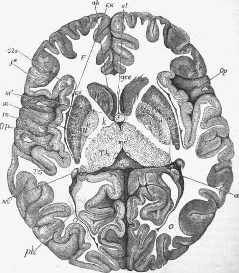
Fig. 321. - Horizontal section of brain of a child nine months old, the right side at a somewhat lower level than the left. F, frontal; TS, temporo-sphenoidal; and 0, occipital lobes; Op, operculum; hi, island of Reil; Cls, claustrum; /'", third frontal convolution; Th, thalamus; A'C, nucleus caudatus; NC', tail of same; LN, nucleus lenticularis; /, //, ///, its first, second, and third divisions; IK, internal capsule, posterior division; IK', anterior division, and k, knee; ah and ph, anterior and posterior horns of left lateral ventricle; gcc, knee of corpus callosum; sp, splenium; mc, middle commissure; f, fornix; sl, septum lucidum; a, cornu Ammonis. (Ross and Flkchsio).
Continue to:


My Books

