


IV. Tumours And Parasites Of The Meninges
Description
This section is from the book "A Manual Of Pathology", by Joseph Coats, Lewis K. Sutherland. Also available from Amazon: A Manual Of Pathology.
IV. Tumours And Parasites Of The Meninges
Tumours of the membranes of the brain are of importance especially when they press on the brain or on the nerves as they issue from the skull. They are of considerable variety.
Fibromas have been found arising from the dura mater both of the brain and of the spinal cord. In the latter case they are liable to press on the cord and may even interrupt it. In a case observed by the author a hard fibroma the size of a marble produced serious mischief by pressing on the cervical cord.
The Chondroma has occasionally been observed, especially in the dura mater of the cord. In a dog observed by the author, paraplegia was produced by such a tumour in the dorsal region.
The Lipoma is a very rare form of tumour in the meninges; that already mentioned on the surface of the corpus callosum (p. 214) probably originated in the pia mater.
The Psammoma occurs not infrequently in the dura mater, where it forms a rounded growth on its internal surface (see Fig. 348); sometimes it is multiple. The tumour is usually small, but may be as large as a walnut, and its surface is rough and tuberculated. The psammoma also occurs as a small soft tumour in the choroid plexus and in "the pineal gland. In the latter case there may be a tumour of considerable size, whose structure is like that of the normal gland, and so the condition is sometimes designated Hyperplasia of the pineal gland. There may also be a Papilloma arising from the choroid plexus either in the lateral ventricles or the fourth ventricle.
The Osteoma hardly occurs in the membranes as a distinct tumour but it is common to find flat pieces of bone in the dura mater, especially in the falx and tentorium, and even in the arachnoid.
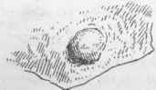
Fig. 348. - A small psammoma of the dura mater, natural size.
The Pacchionian bodies sometimes give origin to tumours which may grow to considerable dimensions. These bodies consist of papillae which spring from the arachnoid and project in various directions; some of them impinge against the skull, where, after thinning the dura mater, they cause pits; others project into the longitudinal sinus.
Cleland has described two tumours of a papillary character which grew from the dura mater and pressed on the brain substance. Although one of these was situated in the falx, which does not normally present Pacchionian bodies, yet the structure of the tumours conformed to that of these bodies. In that on the falx there were calcareous masses like those which characterize the psammoma. The tumours measured respectively two inches and one and a half in diameter.
Sarcomas are exceedingly important tumours, especially those of the Sura mater. Most of them are soft in consistence and contain chiefly round cells. Sometimes there is an alveolar structure, and the cells have a distinct resemblance to epithelium, so that the tumours have been often described as Cancers. There are even tumours remarkably like epitheliomas sometimes observed in the dura mater; they are generally regarded rather as endotheliomas. The sarcomas generally present great malignancy. On the one hand, they extend and involve neighbouring parts of the dura mater as well as the soft membranes and the brain; and, on the other hand, they press outwards on the skull, and may, after destroying the bone, work through to the external surface. These tumours have their seat mostly at the base of the skull and often lead to serious nervous lesions. They may involve directly such structures as the pons or medulla oblongata, the sarcomatous tissue growing into and replacing the proper structure; they also grow into the cranial nerves. By involving the bone and enlarging it, they may impinge on the foramina by which the nerves take exit. As they are seated usually at the base, they generally, when they work through the skull, present in the pharynx or nares.
Of the remaining tumours, the Syphilitic have been already con sidered (p. 679). Tubercular growths hardly ever occur except by propagation from disease of the vertebrae. Dermoid cysts of the dura mater have been observed in a few cases.
The Echinococcus sometimes develops in the neighbourhood of the spinal canal, in the muscles or in the vertebrae. By enlarging it may extend into the canal, pushing the dura mater before it and compressing the cord. It has even been found to have its seat inside the dura mater. A few cases of Cysticercus in the subarachnoid space have been described.
Literature
Beigel, (Cysts of choroid plexus) Path, trans., 1869, xx.; Kelly, (Papilloma of 4th vent.) do., 1873, xxiv.; Cleland, Glasg. Med. Jour., xi., 1861; Davaine, Traite des entozoaires, 1878; Mader, (Cysticercus in 3rd vent.) Berichte des Rudolfstiftes in Wien, 1872; Gowers and Horsley, Tumour of cord, Trans. Med. Chir. Soc, Lond., lxxi.
Continue to:


My Books

