


Leukaemia. Leucocythaemia
Description
This section is from the book "A Manual Of Pathology", by Joseph Coats, Lewis K. Sutherland. Also available from Amazon: A Manual Of Pathology.
Leukaemia. Leucocythaemia
These terms mean literally white blood and white-cell blood, and they express a condition in which the blood is light in colour, from the fact that the white corpuscles are in great excess. The excess of leucocytes is expressed by the term Leucocythaemia, originally applied by Bennett, but now generally discarded for Virchow's term Leukaemia. The disease is not, however, fully expressed by saying that the leucocytes are in excess. There is also a great diminution in the red corpuscles, so that in this respect the condition might be classed as an anaemia.
Causation
This is entirely obscure. There have been a few cases in which an injury to the spleen is supposed to have been the starting-point, while others have been ascribed to syphilis, rickets, malaria. The disease presents many features which suggest analogies with infective diseases, and the analogy of the tissue lesions with those in Hodgkin's disease, which belongs to that class, is consistent with this view. Cases have been recorded in which pernicious anaemia developed into leukaemia (Waldstein). The disease is twice as frequent in males as in females.
Character Of The Morbid Changes
The normal proportion of white corpuscles to red in the blood is stated as about 1 in 300 to 1 in 450, but it varies within normal limits. There is, after haemorrhage, as we have seen, a slight increase in the proportion of white corpuscles, a leucocytosis, but it is of no special significance. There is also an excess of white corpuscles in many infectious and infective diseases, as pyaemia, erysipelas, relapsing, typhoid, and intermittent -fevers, etc. In leukaemia, however, the relative proportion of white to red corpuscles is greatly altered. A case is not a very severe one in which the corpuscles are as 1 to 10, and they may be as 1 to 2, or even equal.
The actual number of leucocytes not infrequently reaches 500,000 per cubic millimetre, or more than 50 times the highest normal average.
The blood presents considerable varieties in leukaemia in respect to the characters of the leucocytes present. According to Muir two forms are distinguishable, according as large or small mononucleated cells predominate.
In a large proportion of cases there is a great excess of large leucocytes with oval or kidney-shaped nuclei. These cells may contain neutrophil granules (which are not normal in the mononucleated leucocytes), or eosinophil granules, or even basophil granules. The multinucleated leucocytes are also increased, and nucleated red corpuscles are visible. As large mononucleated cells, and especially eosinophil cells, are abundant in the bone-marrow, this form of leukaemia has been regarded as primarily due to changes in the bone-marrow.
In a smaller proportion of cases the so-called lymphocytes preponderate. There is little or no increase in the multinucleated form, and the large mononucleated corpuscles are scarcely at all present. There are few cells except the lymphocytes, and hence the appearance is much more uniform than in the other variety. It might be supposed that in this form the lymphatic glands would be the chief primary seats of the disease, but there is reason to doubt whether this is always the case (Muir). This form is usually more quickly fatal than the other, the anaemia making more rapid advances. It is an interesting fact that not only does improve ment in the general health lead to diminution in the number of leucocytes, but that acute febrile diseases, such as typhoid fever, miliary tuberculosis, influenza, septic fever, pneumonia, have a similar effect. The reduction is only temporary, and there is a rapid increase at the end of the fever. A similar temporary diminution in the size of the spleen and lymphatic glands occurs.
In leukaemic blood, after death, there are found small, colourless, glancing crystals of an octahedral shape, which are usually called from their discoverer Charcot's crystals (Fig. 21). There are also certain other abnormal chemical constituents, chiefly glutine and hypoxanthine.
The specific gravity of the blood is reduced in consequence of the low specific gravity of the leucocytes, as compared with the red corpuscles. It may fall from 1055 to 1040 or 1035. The blood has not a watery appearance. It is even a thicker fluid than usual, and distinctly more opaque, resembling a mixture of pus and blood.
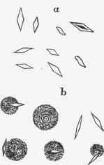
Fig. 21. - Charcot's crystals from the blood in leuksemia after death. In b the crystals are partly inside the white corpuscles. (Zenker.) x 500.
Nature Of The Disease
Seeing that leukaemia is characterized by a great diminution in the red corpuscles and a great increase in the white, the natural inference is that it is due to a delayed or diminished conversion of white into red, and this was Virchow's original view. In the existing obscurity as to the origin of the red corpuscles this view cannot be regarded as established. The disease evidently owes its origin to a grave defect in the formation of the blood, and this is to be associated with primary changes in the blood-forming organs.
Continue to:


My Books

