


Syphilis. Continued
Description
This section is from the book "A Manual Of Pathology", by Joseph Coats, Lewis K. Sutherland. Also available from Amazon: A Manual Of Pathology.
Syphilis. Continued
Tertiary Lesions
These are chiefly characterized by the formation of tumours to which the name G-ummata is applied. They are composed similarly to the indurated chancre, of granulation tissue, but in them the process is much more chronic, and the tissue has a much greater tendency to undergo caseous necrosis; it is also accompanied by a new-formation of connective tissue.
To the naked eye the gumma is a whitish or greyish body, commonly with a yellow caseous appearance in its central parts, or irregularly distributed. It varies in size; it is sometimes as small as a millet seed, in which case it is usually multiple, but it is generally much larger, and may attain the size of an apple. The tumour is not generally sharply denned, but its periphery merges in a firm connective tissue which usually extends outwards into neighbouring structures, so that the tumour appears planted in the midst of a cicatrix.
Under the microscope the tumour will be found, as in Fig. 124, to replace a certain portion of the normal tissue. The central caseous part will be opaque as in c, Fig. 124, Externally the tissue is more transparent (b, Fig. 124), while around and in neighbouring parts of the organ there is new-formed connective tissue, as at d in the figure.
In Fig. 125 the appearances seen under a higher power are shown, the parts taken being from b and c in Fig. 124. It is seen that the peripheral portions (Fig. 125, a) of the tumour present innumerable round cells mixed with fibrous tissue, which latter is often very pronounced. The caseous parts are opaque and present fine fat granules with shrunken cells and nuclei (Fig. 125, b).
The tumours are met with in almost all the tissues of the body, skin, mucous membranes, subcutaneous tissue, in the substance of muscles (as in the tongue), heart, periosteum, liver, dura mater, soft membranes of the brain, cerebral nerves, etc. The name gumma does not express their usual consistence, and is stated to have been first applied to the periosteal form.
The caseous necrosis leads to various results, according to the situation and circumstances of the gumma. If the tumours have a superficial situation, then Ulceration results, and we have a deep excavated ulcer with swollen infiltrated walls, consisting of tissue like that of the gumma, and with the same tendency to degeneration, so that the ulceration extends. As the tumour involves neighbouring structures which undergo necrosis along with the caseous process in the tumour, there may be great destruction of tissue brought about. In internal organs the caseous material may long lie apparently unaltered. The gumma may be virtually healed, its granulation tissue absorbed or converted into connective tissue, while the caseous matter remains, and is finally left in the midst of a cicatrix where it may become calcified.

Fig. 124. - Gumma of liver. Explanation in text. X 16.
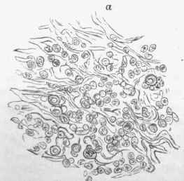
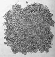
Fig. 12. - From gumma of liver, same section as Fig. 124; a, taken from recent part (b, in other figure); b, from caseous (c). x 350.
We have still to inquire what may be the relation of these tertiary lesions to the virus. The virus no longer exists in the blood, and in accordance with this the lesions are characteristically unsymmetrical. The most probable supposition is that, after the close of the secondary stage, some of the virus has remained lying in a particular part. Perhaps a small gumma has formed, and the virus has lain in it quiescent buff still surviving. It may be waked up by some accidental circumstance in the life of the patient, at any period, afterwards, perhaps as long as twenty years. The virus propagates itself, but its effects are local. It may produce a tumour of large size, but it does not pass into the blood, and therefore does not produce the lesions of the secondary stage. It has been matter of dispute whether a tertiary gumma is an infectious lesion, and-the fact that the blood of the patient himself does not become infected might seem to answer the question in the negative. It is to be remembered, however, that the affected person already possesses an immunity by having passed through the secondary stage. The gumma is probably capable of producing syphilis when its juices are brought into contact with the tissues of a susceptible person.
While the conditions described as characteristic of the tertiary stage usually succeed those of the secondary period, it should be understood that there are great variations both in degree and order of occurrence. The secondary manifestations may be greatly prolonged, and the tertiary may develop to some extent coincidently. The seat of the tertiary lesions varies very greatly.
The tertiary stage of syphilis is often in its later period associated with Amyloid disease. This may be due in some cases to chronic suppurations induced by the specific lesions, but the observations of Fagge show that it is not always so. Of 76 cases of amyloid disease ociated with syphilis there was evidence of former or present bone disease only in 34. In the total autopsies in cases of syphilis over a period of years, amounting to 177 cases, amyloid disease was present in 76 cases or 43 1/2 per cent.
The absorption of a gumma may be promoted by the adminstration of remedies. The exact process by which this is brought about is hardly known, but there seems to be a simple fatty degeneration with absorption, in the way indicated in the section on fatty degeneration.
Syphilis is often associated with a condition of the arteries which will come up for discussion further on. Wherever there is, as so frequently happens, a considerable formation of granulation tissue passing into connective tissue, the arteries take part in the inflammation, and we have, especially, thickening of the internal coat, sometimes going on to complete obliteration of the calibre of the vessel. (See Fig. 126.) This is sometimes very strikingly seen in the neighbourhood of gummata, and by diminishing the blood-supply, it may contribute to the degeneration of the gumma. It may also lead to degenerations in parts around, as where softening of the brain occurs in connection with gummata.

Fig. 125. - Syphilitic affection of meninges and brain, b, b, Arteries showing thickening of internal coats.
Hereditary Syphilis
We have already seen that syphilis, in the secondary stage at least, may be transmitted to the offspring. In the acute period death often occurs in utero, or the child sickens soon after birth and dies within a few weeks. But it often happens that the children do not show any evidence of syphilis for months or years. In this way we may distinguish cases of congenital syphilis from cases of simple hereditary syphilis, the former being born with syphilitic lesions, the latter developing them afterwards.
In Congenital syphilis the most constant and unequivocal lesion is the affection of the bones, which will be considered afterwards. In this condition there is an error in the process of ossification, with inflammatory conditions.
In Hereditary syphilis the lesions are, like those of the secondary stage, mainly inflammatory. There are inflammations of the skin, mucous membranes, cornea, etc. The characteristic malformation of the teeth which Hutchinson has pointed out seems related to inflammation of the mucous membrane of the gums during the development of the teeth.
Literature
The author, in describing the general pathology of syphilis, has followed chiefly Hutchinson and Virchow. Hutchinson, On Syphilis, 1887, and Debate on Syphilis, Path, trans., vol. xxvii., 1876; see also other speakers in this debate; Virchow, Krankhafte Geschwiilste, ii., p. 393; Lang, Path, und Therap. der Syphilis, 1896; Ricord, Traite prat, des malad. ven., 1838; Lancereaux, Traite hist, et pratique de la syphilis, 1866; Hilton Fagge, Medicine, vol. i., p. 109, and Path, trans., vol. xxvii.; Kassowitz, Die Vererbung der Syphilis in Strieker's Med. Tahrb., 1875, p. 359; Fournier, Lecons sur la syphilis, 1881; Van Harlingen, International Encycl. of Surgery, vol. ii., 1882. Bacillus of Syphilis - Lustgarten, Med. Jahrb. d. Wien Gesellsch. der Aerzte, 1885. Since the publication of Lustgarten's observations many have observed the bacillus, but some authors have not found it and others have stated that it exists in the normal smegma of the prepuce. This controversy is chiefly in the pages of the Deutsche med. Wochensch. See also Bitter, Virchow's Arch., cvi., 1886.
Continue to:


My Books

