


Taenia Solium. Continued
Description
This section is from the book "A Manual Of Pathology", by Joseph Coats, Lewis K. Sutherland. Also available from Amazon: A Manual Of Pathology.
Taenia Solium. Continued
As the proglottides become mature they sever their connection with the worm and drop off from its lower extremity one by one. They pass down the alimentary canal, and are discharged with the faeces, or else work their way out through the anus by virtue of their contractile power. For a short time after discharge they still show a writhing movement, but they soon come to rest and die. By the decomposition of the proglottis the ova are set free and are ready under suitable circumstances to develop further.
It is mostly in the bodies of swine that the taenia solium passes through the next phase of its development, although sometimes it occurs in man.
The Ova (Fig. 177) are surrounded by a dense shell of a brownish colour. Inside the shell the egg develops an embryo which acquires six boring spines. When such ova get into the intestinal canal of the pig, the shell bursts, and the embryo with its spines escapes. It proceeds to bore its way outwards, and after piercing the alimentary canal, it finds its way to the muscles of the animal where it finds a lodgment.

Fig. 177. - Ova of T. solium, a, with yolk; b, without yolk, as in mature segments. The hard brown shell is indicated. (Leuckart).
Arrived at its desired seat, the embryo comes to a state of rest, and after a time develops into the scolex, or Bladder-worm, which is, in the case of this species, called the Cysticercus cellulosae. The appearance of these cysticerci in the muscular tissue is shown in Fig. 170, which is drawn of the natural size. The complete cysticercus or scolex is composed of a sac, connected with which is a head, which closely resembles the head of the mature worm, and possesses similar hooklets (Figs. 178 and 179).
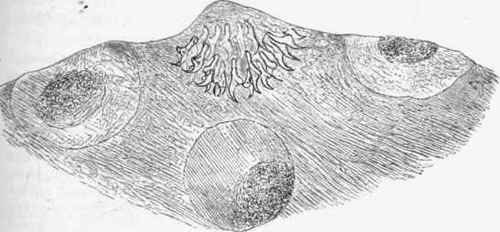
Fig. 178. - Head of cysticercus cellulose;e found in substance of brain, x 90.
The Cysticercus cellulosae occasionally develops in the human subject. It occurs chiefly in the brain, in the eyeball, and in muscle. In rare cases the cysticercus assumes in the brain a very peculiar character. The cyst, developing in the membranes on the surface of the brain, presents pouches and swellings which give it somewhat the character of a bunch of grapes, and so has arisen the designation Cysticercus racemosus. Heads are seldom found in these bunches, and at most there is but one, which has the characters of that of the taenia solium. The cysticercus is usually surrounded by a connective-tissue capsule which is produced from the surrounding tissue and encloses both cyst and head, but not infrequently, especially in the brain and eyeball, it is devoid of this secondary capsule. In that case the vesicle sometimes grows to considerable dimensions, and the head is able to protrude itself and move about in various directions, perhaps in the ventricle of the brain or the eyeball. In these parts the scolex may produce considerable disturbance.
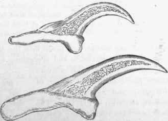
Fig. 179.- Hooks from head of cysticercus cellulosae. x 350.
Although capable of a considerable duration of life, after a time the scolex usually dies, and then it shrinks and becomes, probably, incrusted with lime salts.
The observations of Leuckart and others have thrown much light on the development of the scolex, as they are based on actual experiments in which swine were fed with the ova.
In the first place a vesicle or cyst is formed in the muscle. After a time a slight thickening of the wall of the cyst appears. This grows inwards into the cyst, carrying with it, however, the external wall, so that the projection inwards is hollow with an internal canal continuous with the external surface of the cyst, and so opening externally; this is shown in Fig. 180, where a portion of the wall of the cyst is preserved, and the projection is shown with its internal cavity communicating with the surface of the cyst. This projection enlarges, and by and by the peculiar structures of the head, namely, the four sucking discs and the hooklets, show themselves. But these are formed inside the canal, near its inner extremity, and they are in an inverted position as compared with those of the mature tape-worm. The head with its hooklets is thus at the bottom of the canal, and the four suckers, looking towards each other, follow. After a time the head acquires the power of inverting itself outwards, and thus projecting from the vesicle, or again withdrawing itself within the vesicle as before. This is effected by means of muscular fibres. For the completion of this phase of development a period of from three to four months is required from the time of the ova being taken into the alimentary canal.
When living scolices are taken into the alimentary canal of man, in the first place, the vesicle and every thing, but the head and neck are lost, and we have a small creature which has considerable power of elongating and moving about its suckers, as shown in Fig. 181, The head now fixes itself to the wall of the alimentary canal, and the body begins to develop from its posterior extremity. It takes eleven or twelve weeks for the worm to assume its full dimensions, and at the end of that time it begins to shed proglottides. The worm is of tolerably long life, and may inhabit the intestine of its host for many years. It not infrequently happens that several co-exist in the same person; as many as 30 or 40 have been observed.

Fig. 180. - Cysticercus with beginning of development of head. A portion of the original cyst is shown with the projection inwards of a hollow process which communicates externally, x 25. (Leuckart).
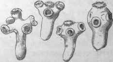
Fig. 181. - A single head of taenia solium before segmentation has begun. It shows movements of its suckers, etc. x25. (Leuckart).
Continue to:


My Books

