


Tuberculosis. Part 5
Description
This section is from the book "A Manual Of Pathology", by Joseph Coats, Lewis K. Sutherland. Also available from Amazon: A Manual Of Pathology.
Tuberculosis. Part 5
Effects Of Local Tuberculosis
The process of tuberculosis, as already described, involves destruction of tissue, and in many cases ulcers and cavities are the result. The destruction of tissue may itself involve serious consequences, as in the case of the bones and of the brain, in which latter position the tubercular mass also acts as a, tumour, pressing on the brain substance around. Most of the evil consequences, however, arise in connection with ulceration and formation of cavities. The surfaces thus produced discharge and use up the available nutritious material of the body. Such processes are, therefore, accompanied by Emaciation. Even more potent in the production of general emaciation is Fever, which commonly accompanies tuberculosis. Fever in general is produced by the presence of foreign matter in the blood (see further on). In tuberculosis the bacilli do not produce fever directly, as they are not present in the blood, but their products are, in a dilute form. In the case of discharging surfaces, again, there will commonly be putrid and other forms of decomposition in the matter discharged, and the absorption of the products will still further conduce to fever.
General Tuberculosis
This term is used in contradistinction to local tuberculosis, to designate a condition in which the tuberculosis has not one but many centres to which the virus has been carried by the blood. It has been pointed out above that tuberculosis has little tendency to penetrate deeply, preferring to extend along open channels or surfaces. Hence, the disease, for the most part, remains local. When conveyed into the blood, the bacilli may or may not settle •down and multiply. This will depend on the number and vigour of the bacilli, and the resisting power of the tissues. If they are present in large numbers, as where a local lesion bursts into a blood-vessel, then they are planted in many organs, and produce their usual effects, namely, the formation of tubercles and inflammation. Hence, in general tuberculosis we have what is equivalent to a multitude of local tuberculoses.
For the most part the disease is a very acute one, hence the names Acute general tuberculosis and Acute miliary tuberculosis. There is a simultaneous and wide-spread eruption of tubercles in several organs. The tubercles have the usual structure, but they are present in vast numbers in the lungs (see Fig. 131), liver, spleen, kidneys, sometimes in the meninges, lymphatic glands, supra-renal capsules, etc. The author has seen them in the heart-wall and endocardium. According to Tripier the bacilli may settle on the valves and produce an acute tubercular endocarditis, in his case of the mitral valve. The tubercles-often show their relation to the circulation by their connection with the blood-vessels. In the lungs they are very frequently grouped around the smaller arteries, and may even form in the internal wall of the artery, projecting into its calibre. This is shown in Figs. 131 and 132, and such an appearance is not uncommon. The disease is an acute febrile one, fatal in a few weeks, and the individual tubercles are small in size, each local formation being little more than a single miliary tubercle. They are often so small as to be only detectable by the aid of the microscope, especially in the liver, where they are present in almost incredible numbers.
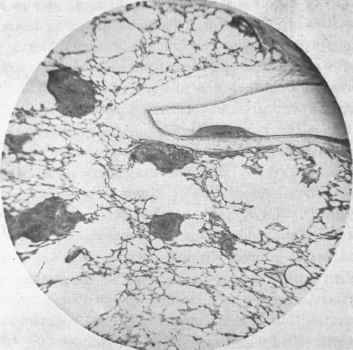
Fig. 131. - Photograph of a section of lung in a case of acute general tuberculosis. A tubercle has formed inside an artery.
The simultaneous outbreak of numerous tubercular lesions implies that in a short interval the blood has received large numbers of bacilli. In some cases the bacilli have been detected in the blood during life (Weichselbaum), and not infrequently after death.
This overloading of the blood with bacilli can only occur when tuberculosis has extended so as to involve the wall of a still pervious vessel of some size, so that the bacilli may find free access to the blood. Acute general tuberculosis has been found associated with tuberculosis of the thoracic duct, of the pulmonary, splenic, portal, hepatic, and other veins. The extension may also occur from lymphatic glands directly into the blood-vessels of these glands (Koch).

Fig. 132. - Photograph of a tubercle in the wall of a small artery in acute general tuberculosis. Two giant-cells in the midst of the tubercle. X ICO.
This solution of the problem of acute general tuberculosis was first suggested by Ponfick, who observed tuberculosis of the thoracic duct in a case of this disease. Weigert has, in a large proportion of cases, found the source of the general infection in the extension to pervious veins, and he asserts that in most cases such a source will be found if diligent search be made. The pulmonary vein is that most frequently involved, and it is so in about half the cases. In a case observed by the author, in which the primary tuberculosis affected the lung, a branch of the pulmonary vein, the size of a crow quill, presented in its wall an elongated, yellow, caseous layer, somewhat resembling a thin localized thrombus. The surface was smooth and there was no layer of fibrine between it and the calibre of the vessel which was here uninterrupted, except by the slight projection of the yellow mass. Tubercle bacilli were found in large numbers in this yellow structure, extending to its internal aspect. Bacilli were also found in the blood and in the miliary tubercles in lungs, liver, etc.
Literature
General Works - Bayle, Recherches sur la phtisie pulmonaire, 1810; L.ennec, Traitede l'auscult. med., etc., 1819; Waldenburg, Tuberkulose, etc., 1869; Virchow, Geschwulste, vol. ii., 1864-65; Discussion on pulmonary phthisis in relation to tubercle of lung, Path. Soc. trans., 1873; Cornet, Die Tuberculose, 1899. Inoculation and Tubercle bacillus - Villemin, Du tubercle au point de vue de son siege, etc., 1862; and Etudes sur la tuberculose, 1868; Langhans, Die Ueber-tragung der Tuberk., 1868; Klebs, Virch. Archiv, vols, xliv., xlix., etc.; Tappeiner, Virch. Archiv, vols, lxxiv. and lxxxii.; Cohnheim, Die Tuberkulose vom Stand-punkt der Infectionslehre, 1880; Sanderson, Researches on artificial tuberculosis, 1869; Koch, The Etiology of tuberculosis, Syd. Soc. trans., 1886; Baumgarten, Many papers, and Tuberkel und tuberkulose, 1885; Weichselbaum, Deutsche med. Wochenschr., 1884; Johne, Die Geschichte d. Tuberkulose, 1883; Karg (Postmortem tubercles), Centralbl. f. Chir., 1885, No. 32. Anatomy and mode of extension - Vikchow, Geschwiilste, vol. ii., 1864-65; Kindfleisch, Path. Histology, Syd. Soc. transl., 1872; Langhans, Virch. Archiv, vol. xlii.; Schuppel, Lymph-driisentuberkulose, 1871; Buhl, Lungenentziindung, etc., 1872; Koster, Virch. Arch., vol. xlviii.; Baumgarten, Tuberkel und Tuberkulose, 1885; Hamiltox, On the path, of bronchitis, etc., 1883; Friedlander, Volkmann's klin. Vort., No. 64; Guterbock (Lupus), Virch. Arch., vol. liii. General Tuberculosis - Ponfick, Berl. klin. Wochenschr., 1877; Zahn, Virch. Arch., vol. lxxvi., 1879; Weigert, ibid., vol. lxxvii., and Deutsch. med. Woch., 1883 and 1885; Tripier, Arch, de med. experimentale, 1890.
Continue to:


My Books

