


XI. Tumours And Parasites Of The Lungs
Description
This section is from the book "A Manual Of Pathology", by Joseph Coats, Lewis K. Sutherland. Also available from Amazon: A Manual Of Pathology.
XI. Tumours And Parasites Of The Lungs
Primary tumours of the lungs are infrequent. Hebbing relates a case of primary tumour replacing the left lung and which he calls a Rhabdomyoma, but which contained cartilage, gland-elements, etc., as well as muscle.
Primary fibroma, osteoma, lipoma, and enchondroma have been observed in the form of small tumours, sometimes multiple. These are unimportant.
Primary canceris a form of tumour which sometimes attains to considerable size. The tumour seems to take origin in the mucous glands of the bronchi, and it usually retains somewhat of the glandular character throughout, presenting in many cases the features of the Cylinder-celled epithelioma. The tumour may, for a time at least, confine itself to the bronchi and their neighbourhood, infiltrating the peribronchial connective tissue. But it often extends to the parenchyma of the lung, forming in some cases bulky tumours. The cancer in the lung has a tendency to soften so as to form Cavities, in whose wall cancerous tissue may be found. In somases most of the cancerous tissue from the primary tumour may be lost by this process of softening, and the case may have the general aspects of a cavity in the lung with an edge in which, even with the microscope, it may he difficult to detect the proper tumour tissue, as it is greatly obscured by inflammatory products.

Fig. 380. - From a primary cancer of the lung, showing exten-rioo by the perivascular lymphatics, a, section of pulmonary artery; b, b, cancerous growth in lymphatics.
In its extension the tumour sometimes penetrates into the lung alveoli. On the other hand, it often forms for itself alveoli similar in size to those of the lung, but of independent origin. It penetrates into the perivascular lymphatic spaces still more than into the lung alveoli, and may often be detected filling up these around the vessels as in Fig. 380. This appearance is sometimes visible a considerable distance in advance of the edge of the tumour.
The cancer sometimes extends to neighbouring structures such as the pericardium, the wall of the heart, etc.
The author has recorded a case in which a very peculiar secondary extension occurred to the bones and to the brain. In these two situations the tumours assumed remarkable cystic tendencies, so that in the brain they were represented chiefly by cysts (see Fig. 338, p. 686). These cysts occurred apparently by mucous or colloid change, although in the primary tumour in the lung there was little such change. The author has observed several other cases in which secondary tumours have occurred in the brain and bones. In some of these the primary tumour in the lung was only represented by a cavity with infiltrated walls, whose nature might have been readily overlooked.
Secondary tumours are somewhat frequent, either by direct extension from the neighbourhood or by embolism.
The lung is often involved by direct extension in Lympho-sarcomas of the mediastinal and bronchial glands. The new-formation follows the connective tissue of the lung, and we find it penetrating in a radiating manner from the root, often burying the bronchial tubes in a sheath of new-formed tissue and partially or completely obstructing them.
Of the tumours arising by embolism, sarcoma and cancer are the chief forms, although chondromas have also been known to extend to the lungs in this way.
Sarcomas, as we know, sometimes penetrate directly into the veins, and portions being carried to the right heart are caught in the branches of the pulmonary artery or capillaries. ' The secondary tumours in the lung are multiple and they repeat exactly the structure of the primary one. Thus we have giant-celled, round-celled, and spindle-celled sarcomas. A tolerably frequent form is the pigmented sarcoma. The En chondroma also not infrequently undergoes secondary development, especially in connection with tumours of the testis.
Cancers, as we have seen, do not readily penetrate into the veins directly. Finding an easier path by the lymphatics they nearly always first develop in the lymphatic glands. After a time the cancerous tissue may penetrate from the lymphatic sinuses in the gland into the veins, or they may possibly penetrate directly from the primary tumour into the latter, and the material is carried on to the lungs. As the cancer is usually arrested for a considerable time at the lymphatic glands, it happens that in all forms of cancer secondary tumours in the lungs are of late development. As the secondary tumours are in the lymphatic glands we may regard those in the lungs as of a tertiary order.
It is clear that the lung is not a congenial situation for the growth of cancerous tumours, as they rarely attain to any considerable size. There is the most marked contrast in this respect between the lungs and the liver. There is not infrequently in the lungs abundant evidence of cancerous infection, but the actual cancerous growth almost limits itself to the lymphatics. Thus there may be visible under the pleura a net-work of a whitish colour consisting of lymphatics occupied by cancerous growth, whilst, on section, small white nodules are seen around the bronchi, which are the peribronchial and perivascular lymphatics filled with cancerous tissue.
The lungs may give passage to the cancerous infection whilst themselves unaffected, or only to the slight extent mentioned above. There may thus be a generalization of the cancer without any definite tumours in the lungs.
An interesting demonstration of the conditions referred to above was afforded in a case which occurred to the author, and which may here be briefly related. It illustrates as well the tendency of the cancerous growth to penetrate into existing spaces and canals. The primary tumour was a cancer of the stomach and secondary growths had occurred in the prevertebral glands. One of these glands was adherent to the vena cava, and several radicles of this vein emerged from the midst of cancerous glands. Actual cancerous thrombi of some of these veins were detected by the naked eye, and under the microscope it was seen that the cancerous masses had partially penetrated into the venous radicles in the glands. On examining the lungs with the naked eye it could be seen from the surface that the new-formation was largely in the lymphatic vessels which were visible as a white network. On section the arteries were usually seen to be surrounded by new-formed tissue. Under the microscope the finer branches of the pulmonary artery were found to be frequently obstructed. The obstructing material was not entirely cancerous in structure, but there was often a round-cell formation with now and again a distinctly cancerous appearance. In most cases the obstruction was complete. Outside the obstructed arteries there were spaces and canals filled with cancerous growths, as shown in Fig. 379. These were obviously the lymphatic channels of the sheath of the vessels into which the cancerous formation had penetrated, just as it does in primary cancer (see Fig. 380).
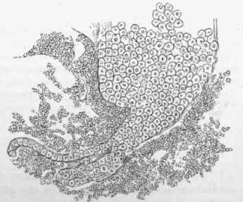
Fig. 381. - Embolic cancer of lung. One of the spaces tilled with epithelial cells. There is a larger space with two brunches into which the epithelial cells have extended, x 90.
Parasites In The Lung
These are of very rare occurrence, unless we include microbes which have already been considered. A fungus of the Aspergillus form has been found in a few cases, in cavities, and in the expectoration in some cases (Mycosis of the lung).
Of animal parasites, Echinococcus is most frequently seen. There may be perforation of the cyst into bronchus, pleura, or peritoneum. The parasite is generally situated in the lower lobe. Cysticercus cellulosse has also been observed.
Amongst rare parasites in the lung may be mentioned one case of a long round worm, the Strongylus longevaginatus, found in a child six years of age (Diesing). It is also said that in Egypt the eggs of distoma haematobium are found in the interstitial tissue. The distoma pulmonale is a frequent parasite in Japan. The worms are about three eighths of an inch in length, and their eggs appear in the sputum of the patients, which also contains blood (see p. 385).
Literature
Tumours - Rokitansky, Handbuch, hi.; Virchow, Geschwulste, ii. Hebbing, Centralb. f. allg. Path., ix., 434, 1898; Ebebth, (Cancer) Virch. Arch., xlix.; Langhans, ibid., liii.; Perls, ibid., lvi.; Weichselbaum, ibid., lxxxv.; Finlay and Parker, Med. chir. trans., Ix.; Marchiafava, Riv. clin. di Bologna, 1874; Coats Path, trans., 1888, xxxix., 326. Parasites. Mycosis - Virchow, Virch. Arch., ix. and x.; Furbringer, ibid., lxvi.; Renon, Iiltude sur Paspergillose, Paris, 1897 (Bibliography). Echinococcua - Thomas, Brit. Med. Jour., 1885, ii., 692; Laveran, Kyst. hydat. des poumons, 1885. Round worms - Manson, Lancet, 1883; Baelz, Berl. klin. Wochenschr., 1883, Die Krankh. d. Athemorgane, Toki'o, 1890; Yamagiwa, Virch. Arch., cxix., 1890.
Continue to:


My Books

