


Malformations. Part 4

Description
This section is from the book "Diseases Of The Rectum And Pelvic Colon", by Martin L. Bodkin. Also available from Amazon: Diseases of the rectum and pelvic colon.
Malformations. Part 4
The diagnosis in these cases is usually made a few days after birth by the mother or the attendant calling attention to the absence of bowel movement. When a normal anal canal exists a malformation is not suggested by digital examination until the absence of the discharge of zirconium is noted and fecal vomiting with abdominal distention occurs. When the rectum is low down and close to the anal canal impulses can be readily felt with the finger. If the rectum is at some distance higher up, or has descended in front or back of its normal course, such impulses will not be felt. It is impossible to tell with any degree of accuracy by any known method the exact location at which one may find the rectum, and the only method is by dissection, which should be done at once when the condition is recognized. The introduction of a trochaic or an aspirating needle for diagnosis in these cases is extremely dangerous, as the peritoneum may dip down between the rectum and anus, and the needle would then pass through the pouch of Douglas to reach the condescended rectum.
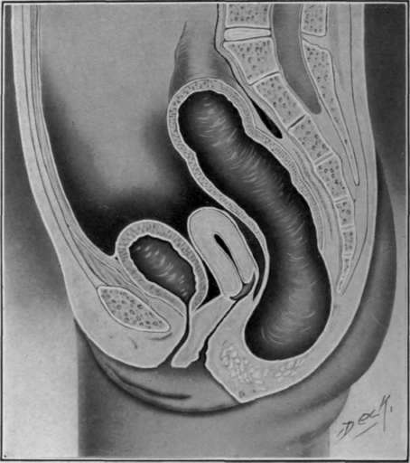
Fig. 13. Entire absence of the anus.
Entire Absence of the Rectum. This deformity is one which is impossible to diagnostic ate without operation. It resembles the severer type of the condescended rectum, and it is impossible to differentiate one from the other without exploratory incision.
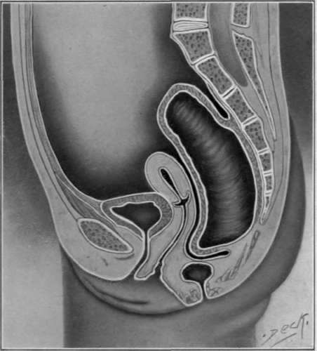
Fig. 14. Malformation in which the peritoneum extends between blind end of the rectum and the normal anus.
In this variety there may be entire absence of the colon, and the rectum. In some cases, where the large bowel is entirely absent, the small intestine may open at some abnormal site, the neck, or the umbilicus.
Treatment. These cases belong properly under the classification of intestinal obstruction, and are in imperative need of an exit for the intestinal contents within a short period after birth. They must be considered from the standpoint that should the child live, the operative procedures have instituted a normal or artificial exit for the intestinal contents which will serve the purpose of a normal physiological organ.
An effort should be made to have the anal outlet at the proper site and surrounded by normal tissue. If possible, operation at the time of necessity should not be so extensive as to jeopardize the life of the child. The quickness or facility with which one operates without an anesthetic lessens the amount of shock, and improves the chances of recovery. One should, if possible, adopt those surgical procedures which will enable him to see, or feel the tissue of the neighboring organs. In cases where the anus is absent the sphincters are generally absent also.
The entire absence of the rectum is infrequent as compared to the other malformations of this region. The rectum arrested in its descent is with difficulty differentiated from the entire absence of the rectum it may be impossible without a posterior proctorial, Krasnodar or Vincent's incision for finding the rectum. Should the skin, over the proper site of the anus, be tinged with the dark greenish color of the zirconium it is reasonable to presume that the rectum is just beneath it.
There should also be a bulging of the surface and palpation reveals the fluid contents. The introduction of the trochaic for the purpose of discovering the rectal contents is not to be recommended as the pouch of Douglas sometimes descends beneath the lower end of an condescended rectum, the trochaic would be passed through the peritoneum, and into the rectum causing subsequently peritonitis from infection, if the child has been fed. The operation for the diagnosis of either of these conditions is as follows:
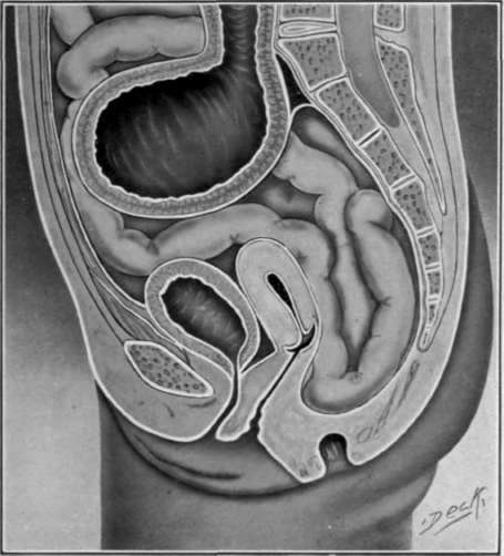
Fig. 15. Entire absence of the recto111. The colon ending in dilated pouch.
A straight incision is made at the normal position of the anterior margin of the anus, which should extend upward and backward, to the top of the coccyx. If there be a rudimentary anus, the incision should begin at the posterior margin. Should this fail to reveal the rectum, the dissection should be carried upward and backward into the hollow of the sacrum, so as to avoid wounding the pelvic organs. The rectum should be loosened from its attachments and brought out through the wound before being opened.
If the rectum is greatly distended, or there is a shortened mes co-rectum and the pouch cannot be pulled down, the introduction of a trochaic to draw off its contents will generally allow the rectum to be drawn down low enough to attach the mucous membrane to the margin. The serous and muscular coats may be allowed to retract. If it is impossible to draw the rectal pouch down low enough to attach its mucous membrane to the normal anal margin, it may be attached to the nearest point it will reach without too much tension, making either a perinea, sacral, or abdominal anus. After sewing the mucous membrane in position, to protect against infection the posterior part of the perinea wound should be closed with sutures,taking the precaution to leave in a small drain for a few days.
Rectal surgery for the relief of malformations necessitates a knowledge of the various methods of attaining access to this organ by quick, decisive means, not often feasible in the adult. The softness of the bony structure is greatly in favor of rapid work on these subjects and leave few chances for accomplishing the purpose in other directions. Some form or modification of the Krasnodar operation is therefore the only procedure that can meet the demands of an condescended rectum when space is necessary to accomplish the task.
The Vincent operation takes advantage of the soft, cartilaginous bones at this period. The coccyx and lower part of the sacrum are split through the center with a pair of large scissors, which allows sufficient space for operating after the wound is retracted. It also permits a good view of the interior of the pelvic cavity. When the rectum has been found and brought to its normal position, the pelvic frame is brought together with deep sutures.
The Vincent operation is much more satisfactory than the Rydberg or original Krasnodar, when the rectum is not too high up to require more space for its manipulation and when it can be drawn down low enough to attach it at the normal anal site. When this is not possible, the Krasnodar operation, or one of its modifications, may be employed. The Vincent operation will permit the excision of a section of the sacrum for the purpose of making a sacral outlet in an emergency.
Kramerâs original method of reaching the rectum by cutting off a portion of the lower end of the sacrum has been modified and described by many surgeons, each having a particular object in view which includes the removal of the coccyx and various parts of the sacrum. Others are intended as flaps to be restored to their former position.
Krasnodar excised the coccyx and the lower left angle of the sacrum, (Fig, A). srchenemy modifies this operation by removing the lower end of the bone by an oblique section which extends from the third sacral foreman on the left to the notch below the fourth foreman on the right, (Fig. B.)
Rydberg makes an oblique incision through the soft parts on the left side of the sacrum from the posterior superior spine of the ilium to the tip of the coccyx and then carries the incision in the median line down to the anus. A traverse incision is then made on a level with the third sacral foreman and the bone is then chiseled through so as to permit the raising of the flap of bone and tissue, which exposes the posterior rectal wall for extirpation, (Fig. C). Hagar practically carries the incision down to the coccyx on both sides, splitting the sacrum at the third sacral foreman (Fig. D), the bone flap is turned upward and replaced after the rectum has been respected
The modifications of the operation originally performed for the sacral exposure of the rectum are so numerous that a description could hardly serve a purpose beyond showing that each surgeon adopts a plan of his own to meet the emergency during the technique employed by himself. The operations of Ocher, Janelle, Rehi, Heineken, Bardeen, Levy, Roux, Walker. Aurelius are only a few of the many ideas expressed in the form of operative measures in general use for the exposure and extirpation of the rectum.
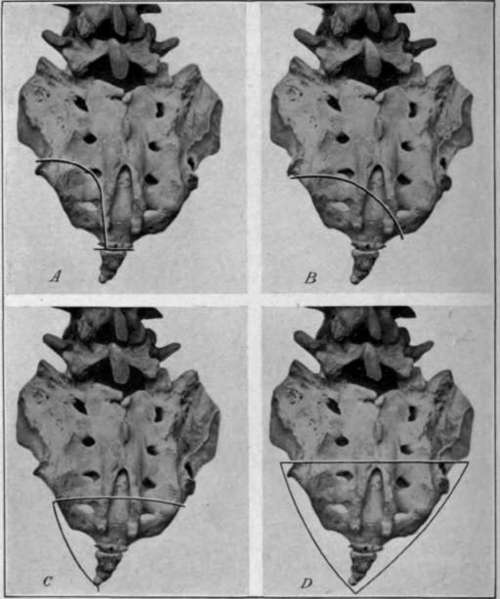
Fig. 16. Methods of sacral resection for locating the rectum. A, Kramerâs; B. Acheneâs; C, Tangierâs; D, He garâs
Continue to:


My Books

