Incorporation Pattern Of Radioactive Estrogens
Description
This section is from the book "The Scientific Contributions Of The Ben May Laboratory For Cancer Research", by The University of Chicago. Also available from Amazon: The Scientific Contributions Of The Ben May Laboratory For Cancer Research.
Incorporation Pattern Of Radioactive Estrogens
The first question we wished to answer with our radioactive hormone was how much actually goes to the tissues which grow. In the young rat, a single dose of estradiol which gives a well-defined but not maximal uterine growth response is 0.1 µg.; so we set out to determine the amount of radioactivity present in various organs and tissues at different times intervals after the administration of this dose by both intravenous and subcutaneous routes. To do this it was necessary to develop a method for the routine assay of tritium in tissues and blood; this assay works well and a detailed report will appear in the literature soon (8).

Fig. 3. Radioactivity in various paper chromatogram fractions on paper chromatography (Bush B-3) of aliquots of high-level stock solution. Fraction I = origin; Fraction IV = estradiol.
Following the single intravenous administration of 0.1 pg. of estradiol (in this case with specific activity of about 9 mc./mg.) to 24-day-old female rats, a maximum of radioactivity was observed in the uterus and cervix between IS minutes and 1 hour. The amount present represents 0.2 % of that administered or 200 µµg. (Fig. 4). Vagina showed a similar pattern with about half as much incorporation, but ovary obtained its maximum radioactivity immediately, with more rapid decrease thereafter. The same relationship in incorporation patterns of uterus, vagina, and ovary following intravenous estradiol were observed repeatedly. When ten times as much hormone (1.0 pg.) was administered, about five times the radioactivity was present in the uterus in the early stages, but rapid clearance of the excess hormone took place, so that after 2 hours about 1.5 as much activity was present with the larger dose (Fig. 5).
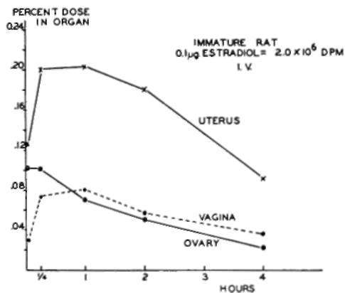
Fjg. 4. Radioactivity in various organs after single intravenous administration of me dium-Ievel estradiol. Each point is the median value for samples from six animals. Uterus includes cervix.

Fig. 5. Radioactivity in uterus after single intravenous administration of different doses of medium-level estradiol and high-level estrone. Each point is the median value of samples from six animals.
A roughly similar pattern was observed in the rat uterus when more highly radioactive estradiol was administered in doses of the order of 0.1 µg. (Fig. 6). After subcutaneous injection the maximum radioactivity was present in the uterus at 15 minutes to 2 hours, corresponding to about 0.15% of the amount injected, or 150 pug. of steroid. After 6 hours this activity had declined to about 0.08 % of the amount administered, or 80 µµg. After intravenous administration the initial radioactivity in the uterus was somewhat higher, but the level soon fell to an amount rather similar to that present after subcutaneous injection.
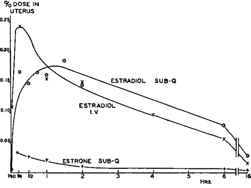
Fig. 6. Radioactivity in immature rat uterus after single injection of steroid in saline. Intravenous (IV.) estradiol: 0.094 ug. (specific activity 96µc./(µg.); subcutaneous (Sub-Q) estradiol: 0.098 ug. (specific activity 117µc./µg.); subcutaneous estrone: 0.10 µg. (specific activity 13µc./µg.). Points for intravenous injections are median values for eight animals; points for subcutaneous injections, for six animals.
Similar orders of magnitude for the uterine radioactivity were observed when 0.05-pg. doses of estradiol in sesame oil were injected daily for 7 days according to the usual bioassay procedure. Twenty-four hours after the last injection, the radioactivity in the uterus was 0.07 % of the single dose (or 0.01 % of the total dose), equivalent to 35 pug. of steroid. Thus, it would appear that, whether administered intravenously or subcutaneously, the amount of steroid present in the uterus during the period when growth is being induced is a very small fraction of the already small administered dose, and is of the order of 30-150 µµg. These amounts are even smaller than those found by Miihlbock (10) and by Emmens (2) to stimulate ramification when applied directly on the vaginal epithelium.
It is interesting to compare the incorporation patterns of uterus and vagina with those of tissues which do not exhibit the marked growth response to estrogens. In Fig. 7, the data from the foregoing single subcutaneous injection experiment are plotted in terms of radioactivity per milligram of dry tissue; since for most tissues 1 mg. dry weight is equivalent to about 5 µl. of fresh tissue volume, the values for blood are expressed in terms of DPM per 5 µl.. Two facts are clear from these measurements. First, the incorporation patterns of liver, kidney, muscle, and blood (and for adrenal and bone not illustrated) are different from those of uterus and vagina, in that the former tissues reach their maximum activity very early with rapid decrease thereafter, whereas uterus and vagina continue to incorporate and retain steroid for a much longer period. Second, with the exception of muscle, the concentration of steroid in the tissues is considerably higher than that in the blood, although, the decrease in radioactivity in all tissues except uterus and vagina appears to parallel the fall in blood level.
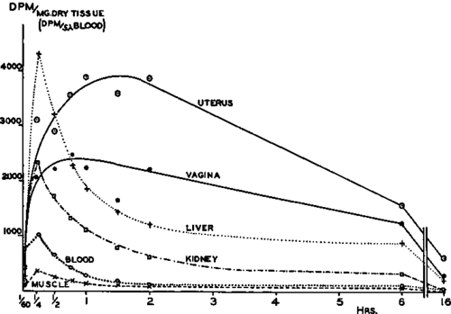
Fig. 7. Concentration of radioactivity in rat tissues after single subcutaneous injection of 0.098 ug. of estradiol (specific activity 117 µc./µg.) in saline. Liver and kidney points are mean values of 3 aliquots of dried pooled tissue; other points are median values of individual samples from six animals.
The difference in steroid incorporation pattern of uterus and vagina from that of the other tissues is more pronounced when lower doses of estradiol are given (Fig. 8). Following a single subcutaneous dose of 0.01 pg. of estradiol in saline, the ability of uterus and vagina to incorporate and retain steroid, when the activity in the other tissues has markedly declined, is quite striking.
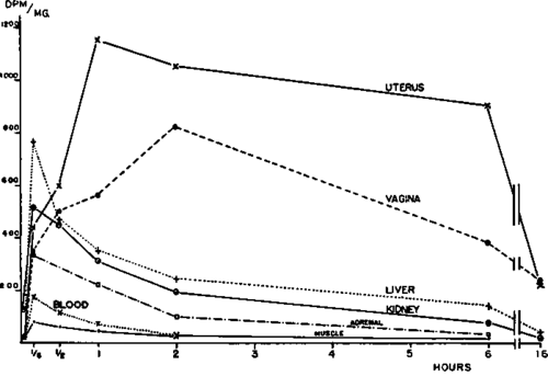
Fig. 8. Concentration of radioactivity in rat tissues after single subcutaneous injection of approximately 0.01 µg. of estradiol (specific activity 195 µc./µg.) in saline. Values are expressed as in Fig. 7. Liver and kidney points are mean values of 4 aliquots of dried pooled tissue; other points are median values of individual samples from six animals.
One other tissue I would like to mention is the pituitary, and here we have done just one experiment so what I say must be taken as only tentative. Following intravenous administration of 0.1 pg. of estradiol, Dr. Sydnor separated the anterior and posterior lobes of the pituitaries which were then assayed for radioactivity (Fig. 9). In contrast to the previous graphs, where each point represents the median value of samples from six or eight animals assayed separately, in the case of pituitary, eight lobes for each time point were pooled, the pools assayed, and each result divided by eight. The anterior pituitary, which is known to be influenced by steroid hormones, shows an incorporation pattern quite similar to that of uterus and vagina reaching a maximum radioactivity which corresponds to 2.5 µµg. of estradiol per pituitary. The posterior lobe, on the other hand, which is not known to be concerned with steroid hormones, showed maximum radioactivity immediately with rapid decrease thereafter. If this preliminary observation can be repeated and confirmed, I think it demonstrates an interesting difference between the two portions of the hypophysis.
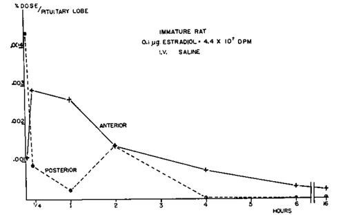
Fig. 9. Radioactivity in different pituitary lobes after single intravenous administration of high-level estradiol. Each point was calculated from single assay of eight pooled organs.
In examining these incorporation curves of estradiol into rat uterus, we wondered whether such studies could furnish information concerning the relation of estrogen structure and activity. It has always been an interesting question as to why it takes ten times as much estrone as estradiol to bring about the same uterine growth response. After intravenous administration of a physiological dose of estrone (1.0 µg.), there was only slightly more radioactivity in the uterus at 2 and 6 hours than from 0.1 pg. of estradiol (Fig. 5); this might suggest that estrone is only one-tenth as efficient in getting into the uterus, if it were not for the fact that administration of 1.0 ug. of estradiol did not give rise to much more activity in uterus during this period. When 0.1 pg. of estrone was administered subcutaneously, however, the amount of radioactivity reaching the uterus was only about one-tenth as much when the same amount of estradiol was given (Fig. 6). If this observation can be repeated and confirmed under different conditions of dose and administration, it may serve to explain the lower physiological activity of estrone as compared to estradiol.
Continue to:
My Books

