Bone
Description
This section is from "The American Cyclopaedia", by George Ripley And Charles A. Dana. Also available from Amazon: The New American Cyclopædia. 16 volumes complete..
Bone
Bone, the substance which forms the internal skeleton of man and the- vertebrated animals, constituting the framework of support, the levers by which force is exerted and locomotion performed, and the boxes or cages in which are enclosed the internal organs. The bony parts of the vertebrated animals are very different in structure and composition from the hard external skeletons of the invertebrata. Bone consists of an organic and an inorganic material, which may be obtained separately by the following simple processes: steep a bone in dilute muriatic or nitric acid; the inorganic or earthy matter is dissolved out, and the organic substance remains, retaining the original size of the bone, and easily bent. In this way is obtained the cartilaginous basis of the bone, on which its shape depends. On the contrary, if a bone be subjected to a strong heat, the organic or animal part is burned out, and the earthy part remains, retaining its form, but crumbling to pieces at the least touch. To the earthy part, which consists principally of phosphate and carbonate of lime, 51 per cent, of the former and 11 per cent, of the latter, the bone owes its hardness, density, rigidness, and white color; to the animal part, principally cartilage, or some form of gelatine, about 32 per cent., it owes its strength of cohesion.
These proportions vary at different ages: in the child, the animal matter forms nearly one half of the bone, accounting for its greater flexibility and the less liability to fracture at this age; in the old, the earthy matter is about 84 per cent., explaining the great brittleness and easy fracture of the bones in aged persons. In the disease called rickets, common among the ill-fed children of the poor in Europe, but somewhat rare in America, there is a deficiency in the deposit of earthy matter, rendering the bones so flexible that they may be bent almost like wax. The power of bone to resist decomposition is remarkable: fossil bones deposited in the ground before the appearance of man upon the earth have been found by Cuvier exhibiting a considerable cartilaginous portion; the jaw of the Cambridge mastodon was found by Dr. 0. T. Jackson to contain 42.6 per cent, of animal matter, and cartilage obtained from the same specimen by means of dilute acid was readily converted into gelatine, and made a good glue; a portion of one of the vertebral spines of Dr. J. 0. Warren's mastodon was found to contain 30 per cent, of animal matter.
The chemical constitution of bone is shown in the following analyses by Berzelius and Marchand:
1.Organic or animal matter........... | 33.30 | 33.25 | |
2. Inorganic or earthy -constituents. | Phosphate of lime..... | 51.04 | 52.26 |
Carbornate of lime...... | 11.30 | 10.21 | |
Fluoride of calcium...... | 2.00 | 1.00 | |
Phosphate of magnesia...... | 1.16 | 1.05 | |
Soda and chloride of sodium... | 1.20 | 1.17 | |
Oxide of iron and manganese, and Loss.... | 10.5 | ||
100.00 | 100.00 | ||
Some recent authorities deny the existence of fluoride of calcium in bone. Bones are not solid. Make a section of almost any bone, and two kinds of structure are seen: one dense, firm, and compact, on the exterior surface; the other loose, spongy, enclosing cells or spaces communicating freely with each other, in the interior of the bone, and surrounded by the more compact tissue. The loose structure abounds in the ends of bones, securing at the same time greater lightness and sufficient expansion to form the joints, while in the shaft or central portion, where strength is most needed, the compact tissue is more developed. Bones are of different forms, according to the uses to which they are to be applied: some are long, as in the limbs, and these are the principal levers of the body; others are flat and thin, composed of two layers of compact tissue, with an intervening cellular structure, destined to enclose cavities. Bones have also a variety of eminences and depressions, for the attachment of muscles, the protection of nerves and vessels, etc.; these eminences, or processes, are well marked in proportion to the muscularity of the subject.
In females and feeble men the bones are light, thin, and smooth, while in the powerfully muscular frame the bone is dense and heavy, and every prominence is well developed. Exercise is as necessary to the strength of a bone as it is to the strength of a muscle; if a limb be disused from paralysis, or the body be prostrated by long disease, the bones waste as well as the soft parts. The external surface is perforated by numerous minute openings, which transmit the arteries and veins to the interior; this surface is covered by a firm tough membrane, the periosteum, composed of densely interwoven white fibrous tissue. The cells, or cancelli, of the spongy portions of bone, are made up of thin and inosculating plates of osseous tissue, enclosing spaces between them which are filled with marrow or medulla; these are lined with a delicate membrane. On a superficial observation it appears as if the plates of the cancellated structure were arranged without definite plan; but the researches of Prof. Jeffries "Wy-man and others show that the cancelli of such bones as aid in supporting the weight of the body are arranged either in the direction of that weight, or in such a manner as to support and brace those cancelli which are in that direction.
The arrangement of these bony plates in the lumbar vertebrae, the neck of the thigh bone, the tibia, and the ankle and heel, is of itself enough to indicate that man, alone of animals, naturally assumes an erect position. This relation is most evident in the above-mentioned bones, and in the adult, it being less observable in youth and old age. There is no real difference between the compact and the spongy structure of bone, the degree of condensation being the only distinction. The cells of the cancelli communicate freely with each other. In the long bones the marrow is not contained in cells, but in one central medullary canal, lined by a membrane. Both the periosteum and the medullary membrane are abundantly supplied with blood vessels, and are therefore intimately connected with the nutrition of the bone, and their destruction to any great extent leads to the death of the part in contact with them. - Microscopic examination can alone explain the intimate structure of bone. If a thin transverse section of a long bone, as the femur, be examined under the microscope, the compact tissue will present several dark circular or oval spots, surrounded by numerous concentric lines; in these lines will be perceived minute black spots, with other lines leading from them in various directions.
The larger oval or circular spots are the openings of vascular canals, called "Haversian," from their discoverer, Clopton Havers; these canals are numerous, taking a course parallel to the axis of the bone, joined together by free inosculation of short transverse branches; they thus form a network of tubes for the minute vessels which they convey and protect. According to Todd and Bowman, the arteries and veins usually occupy distinct Haversian canals, a single vessel being distributed to each. The canals conveying the veins are said to be the larger, and to present at irregular intervals, where two or more branches meet, pouch-like sinuses which serve as reservoirs to delay the escape of the blood; in some of the irregular bones, as in those of the skull, the venous canals are extremely tortuous, running chiefly in the cancellated structure, there called diploe. The Haversian canals vary in diameter from 1/200 to 1/2500 of an inch, the average being about 1/500, and their ordinary distance from each other about 1/120 of an inch.
This whole apparatus of canals is only an involution of the surface of the bone, that the vessels may come into a more free contact with it; as they communicate internally with the medullary cavity, externally with the periosteal surface, and also with the cancellar medullary cells, the network of nutrient vessels is very complete. But, as if this arrangement were not enough to secure the nourishment of such a hard tissue as bone, and so far removed from immediate contact with blood' vessels, there is a still more curious and delicate apparatus of microscopic cavities. Around the Haversian canals will be noticed the appearance of delicate lamellm of bone, more or less concentric; these, with the lacunae mentioned below, are the most essential constituents of true and fully developed bone, the medullary cells and Haversian canals being merely definite spaces existing between the lamellae. It is principally by the successive development of new lamellae that bones increase in diameter, being usually deposited in the direction of the axis.
A transverse section, therefore, would present under the microscope the following arrangement of lamellae, as given by Hassall: 1, several layers passing entirely round the bone; 2, others encircling each Haversian canal; and 3, irregular and incomplete lamellae occupying the angular spaces between those concentrically arranged. The lamellae of the Haversian canals, however, are not exactly concentric, as commonly described, but incomplete and running into one another at various points, a necessary consequence of the irregular distribution of the lacunae. The Haversian systems generally run in the direction in which the tissue requires the greatest strength. With the previously mentioned arrangement of the cancellated structure, the Haversian canals more fully display the wonderful adaptation of means to ends, combining mechanical advantages with the best provisions for the nutrition of the tissue. The number of lamellae passing entirely round the bone is generally less than 12, and those encircling each Haversian canal vary from 2 or 3 to more than 12, the smallest canals having the fewest lamellae.
The lamellae, according to the best observers, appear to consist of a delicate network of fibres in sets, the fibres of each set running parallel, but crossing the others obliquely; some have supposed that they are produced by the union of a number of diamond-shaped cells, and not by the crossing of fibres; the first opinion is probably the true one. Distributed through the cancellated and compact portions of bone occur numerous black specks in the lines of the lamellae; these are the lacunce, or bone corpuscles, the most peculiar and characteristic microscopic form to be found in bony tissue. They differ somewhat in form in different animals, but are always more or less flattened, elongated, ovoid bodies, with numerous branches and radiating filaments passing out from them and communicating with those in the adjacent lamellae. In the dried bone the lacunae are empty, owing to the decomposition and shrinking of the soft parts, and the branched lines running out from them appear as minute canals or canaliculi; but in the fresh condition they are both undoubtedly filled with a soft organized substance, forming an interlacing network of bone corpuscles and filaments, destined to absorb nourishment from the blood vessels occupying the Haversian canals.
The bone corpuscles have an average length of 1/1800 of an inch, and they are usually about one half as wide and one eighth as thick. The diameter of the pores, or canaliculi, is from 1/20000 to 1/12000 of an inch. - From the researches of Mr. Tomes and Mr. Quekett it appears that the ultimate structure of bone consists of a congeries of granular particles, deposited in an organized matrix; these granules are often distinctly visible, without any artificial preparation, in the substance of the delicate spicula of the cancelli, varying in size from 1/6000 to 1/14000 of an inch. The periosteum, a dense, fibrous membrane, richly supplied with blood vessels, covers the external surface of all bones, with the exception of their articular extremities. The vessels of bone are supplied from the periosteum, and ramify, as has been seen, through the Haversian canals; in the long bones a large artery penetrates by the nutritious foramen into the medullary cavity, sending branches to the medullary cells, and inosculating with the capillaries from other sources. Nerves have not yet been detected in the interior of bones supplying strictly the osseous structure, but the painfnlness of many diseases of the bones shows that the external and internal vascular surface must be supplied with nerves.
Lymphatics most probably also exist in bone. - At the earliest period of the appearance of a skeleton in the embryo, it consists of a series of cells; these increase in number and density, and are held together by an intercellular substance, thus forming temporary cartilage, which is afterward converted into bone, though not completely so until adult age. Ossification commences at determinate points or centres, the first of which is in the clavicle, and appears during the fourth week; then follow the lower jaw, ribs, femur, humerus, tibia, and upper jaw; the spine and pelvis are late, and the kneepan does not begin to ossify till after birth. There are generally several ossific centres; for instance, in the long bones, one for the shaft, and one for each extremity. The central part of the bone is the diaphysis, and is not united till long after birth to the ends or epiphyses; processes of bone are called apophyses. Ossification generally extends in the intended direction of the chief strength of a bone.
According to Todd and Bowman, the process by which cartilage is converted into bone is as follows: The small nucleated cells, with comparatively large and granular nuclei, are uniformly scattered through a homogeneous intercellular substance; at the points of ossification the cells begin to assume a linear series, running down toward the ossifying surface, and separated from one another by the intercellular substance; the cells are closely applied to one another, and so compressed that even their nuclei seem often to touch; the lowest rows rest in deep, narrow cups of bone, formed by the ossification of the intercellular substance; the cups are gradually converted into closed areola of bone, with their lamelliform walls. During this first stage of the process there are no blood vessels directly concerned. The lamellae of the areolae, or cancelli, become thicker, and include in their substance elongated oval spaces of a roughly granular nature, in other respects resembling lacunae, and considered by these observers as the nuclei of the cells of the temporary cartilage; within the cancelli only a few cells are found, these cavities being chiefly occupied by a new granular substance, resembling a formative blastema, like that out of which all the tissues are evolved; the cells are in apposition with the wall, and sometimes one seems half ossified, and its nucleus about to become a lacuna; these nuclei have now the same direction as the neighboring lacunae; from the blastema the vessels are probably developed and the necessary elements for the growth of the bone.
The cancelli, at first closed cavities, communicate at a subsequent period, and go to form the Haversian systems, a network of vessels becoming developed within them at the same time. The subsequent process of ossification consists essentially in the slow repetition of the above on the entire vascular surface of the bone. The canaliculi begin as irregularities in the margin of the lacunae, and are converted as the tissue becomes consolidated into the branching tubes which have been described above, and are accordingly formed in the ossified substance of the cartilage cells. As to the lacunae, their granular interior seems to be gradually removed, and they become vacuities for the conveyance of the nutrient fluids. Agreeably to this theory of the formation of bone, Todd and Bowman believe that it grows chiefly, by layers formed in succession on its vascular surface, but also in an interstitial manner after being originally deposited. A most important process of growth is constantly going on in cartilage by the multiplication of the cells and the increase in their dimensions; in the long bones this growth is most active in the longitudinal direction.
Bones also increase by the addition of new systems of laminae on their exterior, and by new involutions of the.vascular surface to form new Haversian canals, as has been proved by experiments with madder mixed with the food of animals; the coloring principle of this substance has a remarkable affinity for phosphate of lime, and it affects first the portions of bone in course of formation, or those nearest to the vascular surface. Wherever there is a vascular network in the structure of bone, whether on the periosteal or internal surface, there growth takes place; the exterior increase is strictly analogous to the exogenous mode of growth in plants. A third mode in which bone grows seems to be by the dilatation of the primary cancelli and central Haversian canals; by this enlargement of the interior the strength of the compact exterior is increased without the disadvantage of an increase of weight. - The reparative power of bone is of the greatest importance in surgery. When a bone is broken, blood is effused, with the coagulum of which a semi transparent lymph is subsequently mingled, covering the surfaces of the wounded parts; in the course of two to three weeks this is gradually condensed by an interstitial change, which' converts it into a substance resembling temporary cartilage; ossification takes place in this in a nearly uniform manner, and the whole is transformed in from four to six weeks into a spongy osseous mass which holds the ends of the bone together; this provisional callus, as Dupuytren called it, is gradually absorbed during the succeeding months, while the permanent callus is being deposited between the contiguous surfaces of the compact tissue; the permanent callus has all the characters of new bone.
When this reparative process is interfered with by meddlesome surgery or constitutional disease, the union takes place merely by ligament, constituting sometimes a false joint. - In reptiles and fishes the cancellated structure usually extends throughout the shaft, which is not so well divided into solid bone and medullary cavity as it is in mammalia. Lacunae are highly characteristic of true osseous structure, being never deficient in the minutest parts of the bones of the higher vertebrata, though those of fishes are occasionally destitute of them. The lacunae of birds are longer and narrower than those of mammals, and the canaliculi are remarkably tortuous; in reptiles they are remarkably long and narrow, and in fishes very angular, with few radiations; their size is not in relation to that of the animal, since there is no perceptible difference between their size in the large extinct iguanodon and in the smallest living lizard. From the emarginated and festooned outline often seen on sections of bone, Dr. 'Carpenter, in his "Principles of Human Physiology," expresses the opinion that the older portions of the osseous substance are removed from time to time, and that the irregular outline thus presented by the Haversian spaces is caused by the partial or complete removal of the Haversian system; in their stead newly formed tissue is deposited; this alternate absorption and reproduction takes place at all times of life, though its energy diminishes with the increasing age of the individual. - The complete development of the osseous system characterizes the final stage of the growth of the organism; the vertebral column does not completely ossify in its spinous and transverse processes until the 25th or 30th year; the ossification of the head and the tubercle of the ribs, commencing soon after puberty, is not continued to the body of the bone till some years after; the ossification of some of the cartilages of the sternum is often not completed even in quite advanced age; the bones of the skull are united within a few years after birth.
As long ago as Aristotle's time, the duration of the life of animals was measured by their period of growth. Buf-fon had the same idea, for he says: " The duration of life, to some extent, may be measured by the time of growth." Animals and man grow only until union takes place between the shafts and the ends of the bones; this union occurs in man at the age of 20 years, in the camel at 8, in the horse at 5, in the ox and lion at 4, in the dog at 2, in the cat at 1 1/2, and in the rabbit at 1 year. Recent observations go to show that animals live about five times their period of growth; this would give, according to Flourens, as the age at which man should arrive, if he lived in accordance with the laws of physiology and hygiene, about 100 years; for the camel 40, the horse 25, the ox and the lion 20, the dog 10, the cat about 8, the rabbit 5 years. In an elephant which died at the age of 30 years, the ends of the bones were not united to the shafts, so that it may be confidently asserted that this animal lives more than 150 years.
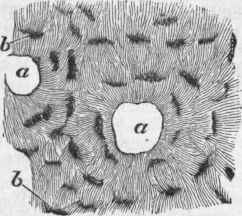
Fig. 1. - Transverse section of bone in the neighborhood of two Haversian canals, a a; b, lacunae.
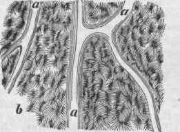
Fig. 2. - Longitudinal section of bone with Haversian canals, a a, and lacunse, b (less magnified than the preceding).
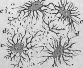
Fig. 8. - Lacunae, c, and canaliculi, d, very highly magnified.
Continue to:
My Books

