Brain
Description
This section is from "The American Cyclopaedia", by George Ripley And Charles A. Dana. Also available from Amazon: The New American Cyclopædia. 16 volumes complete..
Brain
Brain, a collective term, denoting those parts of the nervous system (excluding the nerves) which are contained in the cranial cavity, viz.: the brain, in its popular signification,, or the cerebral hemispheres; the cerebellum, or little brain; and the medulla oblongata, or the upper part of the spinal cord. Each of these has its special part to play in the animal organism. The brain alone, of the animal tissues, is directly influenced by the mental acts of living beings, and through it are effected the mutual reactions of mind and body; the phenomena of sensation and volition, and the mysterious agency of intellect and instinct, are all manifested through the channels of the nervous centres, the most important of which is the brain. The peculiar substance through which all these actions take place exists in two forms, the vesicular and the fibrous. The vesicular nervous matter is gray or ash-colored, granular in texture, containing nucleated nerve vesicles, largely supplied with blood, and is the originator of nervous power; it is sometimes called the " cortical substance," from its forming a thin layer over the exterior of the brain; it is also found in the centre of the spinal cord.
The fibrous nervous matter is generally white, firm, and inelastic, composed of tubular fibres; it is less vascular than the other, and constitutes nearly the whole of the nerves and the greater part of the spinal cord; it simply propagates the impressions sent to or from the vesicular matter. The two kinds do not occur together except in the nervous centres. In the vertebrated animals, nervous matter is a soft and delicate substance, owing the greater part of its tenacity to the vascular and fibrous tissues connected with it. The chemical composition of nervous matter has been well ascertained by Fourcroy, Vau-quelin, and Fremy; but the distinguishing characters of the gray and white substance are as yet imperfectly known. Fourcroy notices the great amount of water in the cerebral matter, from six eighths to seven eighths of its weight, upon which its softness is in great part dependent. According to Yauquelin's analysis in 1812, the brain is an emulsive mixture of albumen, fatty matter, and water holding in solution saline and other matter common to it with other tissues.
The following table gives the result of his analysis:
Albumen..................................... | 7.00 | |
Cerebral fat | stearine, 4.53 | 5.23 |
elaiue, 0.70................... | ||
Phosphorus................................... | 1.50 | |
Osmazome......................... | 1.12 | |
Acids,salts,sulphur.............. | 515 | |
Water......................................... | 80.00 | |
100.00 | ||
The medulla oblongata contains more cerebral fat, but less albumen, osmazome, and water. Fremy's analysis, published in the Annales de Chimie, 1841, confirmed that of Vauquelin, and showed the following proportions: 7 parts of albumen, 5 of fatty matter, and 80 of water. He extracted from the fatty matter the following secondary principles: 1, cerebric acid, a white granular, crystalline substance, containing no sulphur, a little phosphorus, and 66 per cent, of carbon; 2, oleophosphoric acid, separated from the cerebric by its solubility in ether, containing about 2 per cent, of phosphorus in the condition of phosphoric acid, and combined with elaine; 3, cholesterine, the same as that obtained from bile (brains preserved in alcohol are apt to be surrounded by a crystalline substance resembling cholesterine); 4, traces of elaine, margarine, and fatty acids. The brain is remarkable for containing phosphorus, which varies in quantity at different periods of life, being the least in infancy and old age;* the maximum of water is found in infancy, an interesting fact in connection with the serous effusions so prevalent at this period of life; it has been ascertained that the idiot brain contains less phosphorus than the normal organ, this being diminished from nearly 2 to less than 1 per cent., indicating possibly an important hint for the treatment of diseases accompanied by deterioration of the mental powers.
The microscopic elements of nervous tissue are fibres and cells. The fibrous nervous matter, or white central substance, contains tubular fibres or nerve tubes, and the gelatinous fibres found chiefly in the sympathetic system. The white fibres are membranous cylinders, of a pearly lustre, consisting of an external delicate, transparent sheath, within which is a layer of thick, fluid, highly refractive matter, called the " medullary layer; " while the central portion is occupied by a finely granular mass, termed the "axis cylinder." The medullary layer, however, is less distinct in the fibres of the brain than in those of the nervous trunks, and in some instances appears to be altogether wanting. The fibres of the brain average 1/10000 of an inch in diameter, presenting at some points a swollen appearance; they do not communicate with each other like the vessels, nor divide into smaller fibres, but continue unbroken from their origin to their final distribution, inosculating only at their terminal loops. The gelatinous or gray fibres seem to be solid, flattened, transparent filaments, varying in diameter from 1/6000 to 1/4000 of an inch; the mode of their connection with the elements of the nervous centres is unknown.
The essential elements of the vesicular or gray nervous matter are cells, or vesicles, containing nuclei and nucleoli; they are dark, generally globular, but at times very irregular and variously elongated, enclosing a grayish granular substance, and sometimes pigment granules; they vary in size from 1/3500 to 1/300 of an inch in diameter; among the largest of these are the caudate, so called from the irregular tail-like process extending from them.
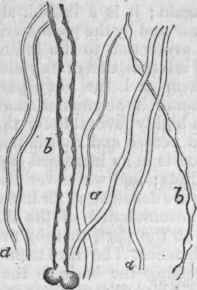
Fig. 1. - Nerve Fibres of the Brain.
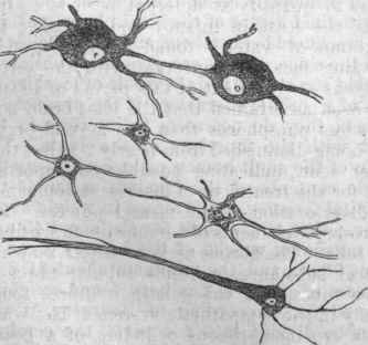
Fig. 2. - Nerve Cells of the Brain.
The nerve vesicles are imbedded in a soft granular matrix in the brain. The nervous centres exhibit the union of these two forms of matter, more widely separated in the brain than in the smaller ganglia; indeed, the cerebral hemispheres are composed internally of fibrous matter exclusively, surrounded by a layer of the gray vesicular substance, into which the fibres are also prolonged. The tubular fibres seem to be capable of regeneration to a certain extent; if the nerve be divided, but the ends not separated, union may take place, and the nerve resume its office-, even when a portion is excised, it appears that true nerve fibres, in smaller number than in the nerve itself, may be developed in the uniting substance, as shown by partial restoration of function and microscopic examination. When a portion of the brain is removed by accident or design, its place is supplied by new substance; but whether this be true cerebral substance or not has not been satisfactorily determined. The white fibres may be distinguished, according to their physiological office, into three kinds - efferent or motor, afferent or sensitive, and commissural or connecting. Henle suggests that there may be a fourth series, associated with the operations of thought.
Of the mode in which the afferent nerves terminate, and the motor nerves commence in the central organs, it may be said that three principal modes have been ascertained, in which there is an actual continuity from one form of nerve tissue to the other: a globular unipolar cell may give out a single prolongation, which becomes a fibre; or a nerve cell may be found in the course of a tube, with each extremity prolonged into a fibre; or some of the radiating prolongations of the caudate cells may become continuous with the axis cylinders of nerve tubes, or inosculate with those of other caudate cells. A curious circumstance in connection with the gray matter is the large quantity of pigment or coloring substance in it, apparently forming one of its essential constituents, as it is everywhere present, though in some situations more abundantly than in others; it has been asserted that this bears a close resemblance to the coloring matter of the blood, and if so, it is a fact of great interest to physicians, who can avail themselves of the restorative properties of iron in cerebral diseases, improving the quality of the nutrient blood by increasing the quantity of the red globules. - The central column or spine of the vertebrate skeleton encloses in its canal the spinal cord; and the cranium, which is a series of modified and expanded vertebrae, protects the continuation of the cord and its expansion into an aggregate of gangliform swellings, the brain or encephalon.
The bruin is enclosed in three membranes, or meninges, continuous with those of the spinal cord, which will be described under that head. From without inward, these membranes are the dura mater, arachnoid, and pia mater. The term mater (![]() , mother) originated with the Arabians, who considered these membranes as the parents of all others in the body. The dura mater is a membrane of white fibrous tissue, strong, flexible, but not elastic; its fibres are arranged on different planes; it is freely supplied with blood vessels, and is perforated for the passage of nerves, and, according to Arnold and Pappenheim, has some branches between its own laminae. It forms the internal periosteum of the skull, and is closely applied to the cranial bones, and in some places firmly adherent, especially in youth and old age. From it processes are given off, which serve as partitions between the cerebrum and cerebellum behind, and between the cerebral and cerebellar hemispheres; these processes are the falx cerebri, which separates the great hemispheres, extending on the median line from the forehead to the occiput, along the sagittal suture; it is falciform in shape, its lower border concave and corresponding to the convexity of the corpus callosum, and its upper border enclosing the great longitudinal sinus; narrow in front, and deep behind, having the inferior longitudinal sinus along its posterior border.
, mother) originated with the Arabians, who considered these membranes as the parents of all others in the body. The dura mater is a membrane of white fibrous tissue, strong, flexible, but not elastic; its fibres are arranged on different planes; it is freely supplied with blood vessels, and is perforated for the passage of nerves, and, according to Arnold and Pappenheim, has some branches between its own laminae. It forms the internal periosteum of the skull, and is closely applied to the cranial bones, and in some places firmly adherent, especially in youth and old age. From it processes are given off, which serve as partitions between the cerebrum and cerebellum behind, and between the cerebral and cerebellar hemispheres; these processes are the falx cerebri, which separates the great hemispheres, extending on the median line from the forehead to the occiput, along the sagittal suture; it is falciform in shape, its lower border concave and corresponding to the convexity of the corpus callosum, and its upper border enclosing the great longitudinal sinus; narrow in front, and deep behind, having the inferior longitudinal sinus along its posterior border.
The tentorium cere-belli extends horizontally between the posterior cerebral lobes and the cerebellum; it is attached to the falx cerebri, and to the occipital and petrous portion of the temporal bones along the groove for the lateral sinus; in the cats and some other leaping animals, this membrane is partially replaced by bone, doubtless to prevent injury from sudden shocks. Between the lobes of the cerebellum descends vertically from the tentorium the falx cerebelli, containing the occipital sinuses. Next to the dura mater, which also furnishes sheaths for the nerves and vessels at their origins, lies the arachnoid, the .serous membrane of the cerebro-spinal cavity; it consists of two layers, the outer one closely adherent to the dura mater, and the inner .one loosely to the pia mater; the space between the two layers is the arachnoid cavity, and that between it and the pia mater, the sub-arachnoid cavity; resembling other serous membranes, the arachnoid is liable to become inflamed, with the effusion of fluid into one or both of the above cavities, especially toward the base of the brain. The sub-arachnoid space is filled with what is called the "cerebro-spinal fluid," varying in quantity from two or three ounces in the healthy adult condition to ten or twelve ounces in old age.
It keeps during life the opposed arachnoid surfaces in contact; it is most abundant where the brain has shrunk either from disease or advanced age. From the experiments of Magendie it appears that its presence is necessary for the healthy action of the nervous centres; when removed, it is quickly formed again; it is a limpid, alkaline fluid, doubtless secreted by the pia mater, and affords mechanical protection to the brain and spinal cord by the interposition of its yielding medium between them and the bony parietes which surround them; its accumulation at the base of the brain is highly favorable for the protection of the large nerves and vessels there situated. This fluid exists in an increased quantity in the brains of idiots; and whenever the cranial or spinal walls are deficient, as for instance in spina bifida, an accumulation of the fluid becomes prominent at the part, thereby protecting the nervous substance. The third membrane immediately investing the brain is the pia mater, composed of white fibrous tissue and blood vessels; in the skull it is very delicate and very vascular; it adheres to the surface of the cerebral and cerebellar hemispheres, and sends innumerable minute vessels to their substance; it sinks into the fissures and sulci, and penetrates into the ventricles, forming the choroid plexuses and the velum iriterpositum; its minute ramifications are sometimes incrusted with sandy particles, consisting principally of phosphate of lime.
The pia mater is the medium of nutrition to the nervous substance and to the arachnoid; and hence any inflammation of these membranes would be communicated to the superficial gray matter of the brain, the seat of its physiological activity. Along each side of the longitudinal sinus it is common to find a series of depressions in the dura mater; these are due to the presence of whitish granules, called Pacchionian glands, from their first describer, of an albuminous material, arising probably from a deposit of granular lymph among the vessels of the pia mater; they are found principally along the edge of the great longitudinal fissure of the hemispheres, pushing the arachnoid before them, and even projecting into the sinus. They are generally considered morbid structures, and the result of local irritation of a chronic character; if the products of disease, they do not seem to interfere in the least with the functions of the brain. - The brain of the adult human male, comprising the whole contents of the cranium as far as the occipital foramen, will average in weight about 50 oz.; that of the adult female, about 45 oz.; the maximum weight of the healthy organ is about 64 oz., and the minimum about 31 oz.; in cases of idiocy it has been found weighing only 20 oz.
According to Bourgery, if the brain be divided into 204 parts, the cerebral hemispheres would weigh 170, the cerebellum 21, and the medulla and sensory ganglia 13; on the same scale, the spinal cord would weigh 7. In proportion to the body's weight, the brain of man would weigh 1/36 part; in the average of mammalia this proportion would be 1/186; in birds, 1/212;in reptiles,1/1321;and in fishes,1/5668 In some apes, rodents, and singing birds the weight of the brain bears a higher proportion to that of the body than it does in man, even as high as 1/12 in the blue-headed titmouse; the increase, however, is not in the cerebrum, the seat of intellect, but in the sensory ganglia, the seat of the instinctive actions. The size of the brain is not in proportion to the physical development of the body; either in animals or man; the horse has a brain inferior in weight to the smallest adult human brain; that of a whale 75 feet long was found to weigh not quite twice as much as that of a man. Even in men there is no fixed relation between the size of the body and of the brain; a small man may have a large brain, and vice versa.
Men of great intellectual power have generally, if not always, possessed large brains; the brain of Cuvier, the great French naturalist, weighed between 59 and GO oz.; that of the French surgeon Dupuytren, 58 oz.; those of Napoleon and Daniel Webster, an ounce or two less. The quality of the brain, however, is quite as important as the quantity, so that a large brain does not of necessity constitute a great man. According to Tiedemann, the female brain, though absolutely smaller than that of the male, is larger when compared with the size of the body. The brain reaches its highest development anatomically at the age of 20 years, which it maintains until 60, after which, in most persons, it begins to decrease in size, with a corresponding decline in the mental powers. There do not appear to be any striking differences between the brains of the various races of man. - For the topographical and pathological anatomy of the brain, an examination from the hemispheres downward is the most practicable method; but for physiological anatomy, it is more advantageous to make the examination from below upward, by which method the student proceeds from the simple to the more complex, following the direction of the fibres of the medulla oblongata to their ultimate distribution in other parts of the brain.
The medulla oblongata is the upper enlarged portion and direct continuation of the spinal cord, extending from the plane of the occipital foramen about an inch upward to the tuber annulare; through this the brain is brought into communication with the other vital organs, and it is therefore the nceud vital, "the link which binds us to life." As its size is proportionate to that of the nerves which proceed from it, it is much larger in some lower animals than in man. Like the spinal cord, it consists essentially of anterior and posterior columns; it may be anatomically distinguished from the cord by the decussation or crossing of some of the anterior fibres. In front are the " anterior pyramids," separated by a median fissure; external to these are the oval protuberances, the " olivary bodies;" more external, and forming the lateral and great part of the posterior portions, are the "restiform bodies," separated from each other in the middle by two slender columns, the " posterior pyramids." The anterior pyramids or fibres extend from the antero-lateral columns of the cord to the cerebral hemispheres, passing through the tuber annulare, the corpora striata, and the optic thalami, contributing to form the lower portion of the crus cerebri; in the tuber annulare these fibres are crossed at right angles by others belonging to it, and are interlaced with them; on tracing them downward, the greater part connect themselves with the middle or lateral columns of the opposite side, while a few are continued down on the same side into the anterior columns of the cord, and others, the "arciform fibres," curve round the olivary bodies and ascend to the cerebellum, not passing to the cord; the anterior pyramids are entirely of a fibrous structure.
The arrangement of these fibres is highly interesting in explaining the phenomena of disease of the brain; since, owing to the decussation of the fibres in the medulla oblongata, an injury of the right side of the brain will produce paralysis on the left side of the body, and vice versa; while a lesion of one lateral half of the spinal cord, below the point of decussation, causes paralysis of the same side of the body. The restiform bodies consist of fibrous strands enclosing a gray nucleus, and pass upward into the crura cerebelli; below they are chiefly continuous with the posterior spinal columns, and partly with the posterior part of the middle columns; as the fibres ascend they diverge, leaving between them the fourth ventricle, and pass into the corresponding hemisphere of the cerebellum, connecting this latter with the spinal cord; the cerebellar columns also communicate by a band of arciform fibres, according to Solly, with the anterior spinal columns; the gray nucleus, or " restiform ganglion," seems to be the ganglionic centre of the pneu-mogastric and a part of the glossopharyngeal nerves.
The posterior pyramids can hardly be distinguished from the restiform bodies externally; but their columns, bounded by the median fissure and by a very slight groove, establish a connection between the sensory tract of the crura cerebri and the posterior columns of the cord; their gray nuclei are the ganglionic centres of the auditory nerves. The olivary bodies, continuous inferiorly with the anterior or motor columns of the cord, and affording attachments to the motor fibres of the first and second cervical nerves, enclose a gray nucleus, and send their fibres forward to the motor tract of the crus cerebri, and backward to the quadrigeminal bodies; the nucleus, or corpus dentatum, seems to be connected with the hypoglossal or motor nerve of the tongue, and also with the glossopharyngeal, one of the sensory nerves of this organ. The medulla is not only a transmitter of fibres from the spinal cord, but is a nervous centre itself; with it are connected the nerves of respiration and deglutition, which are quite independent of the cerebral hemispheres, and beyond the control of the will. - The cerebellum, one eighth of the size of the cerebrum, is placed under the posterior part of the latter, from which it is separated by the tentorium; it is composed of white and gray matter, the former occupying the interior; its convolutions have the form of parallel layers.
Its central part or lobe is the only one found in fishes and reptiles; its lateral lobes, found only in the higher animals and in man, indicate an advance in development. On a vertical section we find the white substance resembling the trunk of a tree from which branches are given off, hence called arbor vita, or tree of life. This' organ is connected with the rest of the brain by three sets of fibres, the superior extending to the tuber-cula quadrigemina, the middle or the restiform fibres passing downward to the medulla, and the inferior or transverse (pons Varolii) passing to the opposite side and forming a considerable part of the tuber annulare; the central lobe has aggregates of lobules on its superior surface, containing both white and gray matter, the "superior vermiform processes," and on the lower surface the " inferior vermiform processes." The transverse diameter of the cerebellum is 3 1/2 to 4 inches, the length 2 to 2 1/2 inches, and its thickness varying from 2 inches in front to less than 1/2 inch behind. For details on the structure and on the intricate divisions of the cerebellum, the reader is referred to special works mentioned at the end of this article.
Disease of the cerebellum, when deep-seated, is generally manifested on the opposite side of the body; this organ presides principally over the regulation of the voluntary movements. The restiform bodies of the medulla in their ascent to the hemispheres of the cerebellum diverge, leaving a lozenge-shaped cavity, the fourth ventricle, bounded above by the median cerebellar lobe, below by the olivary columns, behind by the nodule of the inferior vermiform process, in front by a portion of the superior vermiform process, called the " valve of Vieussens; " on the floor are the white barb-like fibres of the seventh pair of nerves, passing at right angles, and called the calamus scriptorius; it is improperly called the ventricle of the cerebellum, as it belongs to the medulla and is proportionate to it in size. The mesocephalon, or tuber annulare, embraces those portions of the brain which unite the cerebrum above, the cerebellum behind, and the medulla below; the lower surface, or the pons Varolii, consists of curved transverse fibres, passing from one crus cere-belli to the other, crossing apparently over the anterior pyramids like a bridge; they are always developed in proportion to the cerebellar hemispheres, and are absent in animals having only the median lobe; they constitute the great transverse commissure of the cerebellum, as the corpus callosum (mentioned hereafter) constitutes the great transverse commissure of the cerebrum; these fibres extend more than one half of the depth of the tuber annulare.
The tuber annulare, which exists in animals whose cerebellum has no hemispheres, projects from the medulla proper, and contains a nucleus of gray matter; Longet is of opinion that this ganglion is an independent centre of sensation and motor power, and Dr. Todd states that the convulsions excited by a current of electro-magnetism through it are not tetanic, but epileptic, or alternating with relaxations of the muscles. Situated above the tuber annulare are the quadrigeminal bodies, the two anterior being called nates, and the two posterior testes; they are gangliform bodies, containing gray and white matter, the anterior being the larger; these are the analogues of the optic lobes of birds, reptiles, and fishes, in which classes there is only a single pair, but of much larger size. The crura cere-belli, which apparently emerge from the posterior angles of the tuber annulare, derive their fibres from strands going to the testes, from those of the restiform body, and from those of the pons Varolii; from the anterior angles of the tuber annulare diverge two similar processes of considerable thickness, the crura cerebri, which enter the cerebral hemispheres, and upon which each of these masses has been said by Dr. Todd to rest as a "mushroom upon its stalk." The fourth and fifth pairs of nerves are intimately connected with the tuber annulare.
On making a section of the crura cerebri, three planes of nervous matter may be seen: the lower one, of fibrous matter, continuous with the tuber annulare and the anterior pyramids, passes up into the corpora striata, or striated bodies; above this is a dark mass, the locus niger, containing large caudate vesicles abounding in pigment, with nerve fibres among them; the upper layer, of grayish matter, continuous with the central part of the medulla oblongata, or olivary columns, passes up into the optic thalami. The striated bodies and optic thalami are best seen by laying open the lateral ventricles, in which they are placed, closely united to each other, the former being a little in front and outside of the latter. The striated bodies are pear-shaped, tapering gradually backward in a long process which winds down into the anterior extremity of the descending horn of the ventricle, and striated when cut in an oblique direction upward and outward, on account of the passage of the fibres of the crura cerebri into the vesicular matter; through these bodies, by three sets of fibres, communications are established between the tuber annulare, medulla oblongata, and cerebral convolutions; they are generally considered the more essential part of the nervous system which controls voluntary movements.
The optic thalami are of a lighter color, of the same texture and appearance as the olivary columns, of which they are the continuations; a portion projects into the ventricles, and the rest adheres to the striated bodies, the hemispheres, olivary columns, and quadrigem-inal tubercles; the fibres no doubt are continuous with those of the white substance of the hemispheres, and with those of the striated bodies; between them is the third ventricle, the roof of which is formed by the velum interposition, a process of the pia mater. The corpora geniculates, externum and internum, are small gangliform masses, projecting from the posterior part of the optic thalami. Behind the third ventricle is a conical, dark gray body, enclosed by a process of the pia mater, the " pineal body; " it rests in a groove between the nates, and is connected with the thalami by fibres, called peduncles; it consists chiefly of large nucleated vesicles, with a few fibres, and in a cavity near the base contains a sandy substance composed of phosphate and carbonate of lime; its use in the economy is unknown.
The optic thalami have been considered but the principal sensitive centres, without which the sensorium could not perceive the physical change resulting from a sensitive impression; but this is by no means certain. - The cerebral hemispheres constitute the great mass of the brain, and their horizontal section presents an oval, of which the smaller extremity is directly forward; the external surface is smooth, being covered by the arachnoid membrane; they are divided longitudinally along the middle line by the deep fissure which receives the falx cerebri, and at the bottom of which in the middle portion is the great commissure, the corpus callosum; the inferior surface, or base of the brain, is divided into anterior, middle, and posterior lobes, corresponding with the fossa in the cranial bones; the anterior lobe rests chiefly on the roof of the orbits, and on its inferior surface presents the nerve of smell; between it and the middle lobe is the " fissure of Sylvius," through which runs the middle artery of the brain; the middle lobes are gradually lost in the posterior, which are separated from the cerebellum by the tentorium. The space between the middle lobes in the centre is occupied by the pituitary body, crossing of the optic nerves, and the mammillary bodies.
The pituitary body is lodged in the sella turcica of the sphenoid bone, and is a glandiform mass, surrounded by the coronary sinus, and connected with the brain by the infundibular process; it has two lobes, and somewhat resembles the vesicular substance of the brain; its use is unknown. Between the crura of the cerebrum the third pair of nerves emerge. The usual way of examining the hemispheres is to make.a horizontal section at about one third from the summit; this section, denominated the centrum ovale majus, presents a centre of white substance, surrounded by a narrow border of gray, showing the zigzag outlines of the convolutions, and spotted by numerous small red points caused by the escape of blood from the cut ends of minute vessels. In the central line is a broad band of white substance, uniting the hemispheres together as their great commissure, and securing their connected action, the fibres passing from one to the other like a bridge; at its anterior and posterior extremity it is folded downward toward the base of the brain.
On cutting a little deeper, an irrregular cavity is opened on each side the lateral ventricle, containing the striated and optic bodies; these cavities are lined by a serous membrane, secreting a fluid, the undue accumulation of which constitutes hydrocephalus internus, or water on the brain, a fatal disease of children, in which the substance of the brain may become almost obliterated, and the bones of the yet ununited skull distended almost to the size of an adult head. The fifth ventricle is the space between the layers of the septum luci-dum, an extension of fibrous matter connecting the anterior reflection of the corpus callosum with the horizontal fibrous stratum called the fornix, and separating the anterior horns of the lateral ventricles. Between the optic thai-ami and striated bodies in the ventricles, in a superficial groove, is the tcenia semicircularis, a delicate band of fibrous matter, commissural in its character. The posterior horn of the lateral ventricle, according to Owen, is peculiar to man, as also is the hippocampus minor, a projection of one of the convolutions into it; in its inferior horn is the hippocampus major, and a considerable portion of the vascular choroid plexus.
The cerebral hemispheres, after the membranes have been removed, present a peculiar folded arrangement of their surface, the "convolutions;" the folds consist of a layer of gray matter, varying from one eighth to one quarter of an inch in thickness. Physiology has shown that the gray matter of the nervous centres is the originator of nervous force, while the white matter serves only to convey impressions to or from the different parts of the body; hence the greater the number of these convolutions, or, in other words, the greater the amount of the gray substance, the greater will be the physiological power of the brain. In the rat and the mole the surface of the brain is quite smooth; from these the convolutions increase in number up to man. Their arrangement, though never the same in two brains, nor on opposite sides of the same brain, cannot be supposed to be purely accidental; there are certain ones always present (when any exist), whose situation and size influence the disposition of the others; in man the variable and additional convolutions are chiefly on the top and front of the hemispheres. The lower the position of an animal in the scale, and the less developed the organ as we approach infancy, the greater is the symmetry of the two sides.
It is said that the convolutions in the inferior races of man (Todd and Bowman) present a more symmetrical arrangement than is usually found in the more cultivated races. If the gray matter of the cerebral convolutions and the cerebellar layers were spread out, it would occupy about 670 square inches, which by this admirable arrangement are packed into the small extent of the brain. Each convolution consists of a fold of gray matter enclosing a process of the white; the gray matter forms a continuous unbroken sheet over the cerebral surface; the greater part of the white fibres emerge from the gray matter, and thence converge to the central parts of the brain. The fibres which unite different portions of the same or of opposite hemispheres are called "commissures;" the transverse are the corpus callosum, the anterior, posterior, and soft commissures; the longitudinal are the fornix and the superior longitudinal commissure. The corpus callosum connects the great bulk of the hemispheres, especially at the lower part; it is wanting in fishes, reptiles, birds, and the lower mammals. The anterior commissure particularly unites the striated bodies, many of its fibres passing through them and radiating to the middle cerebral lobes; it is very large in the marsupials, which have no corpus callosum.
Thp posterior commissure connects the optic thalami, and is connected with the pineal body. The soft commissure also passes from one optic thalamus to the other, dividing the third ventricle into an upper and lower portion; unlike the other commissures, it contains gray matter. The superior longitudinal commissure is enclosed in the convolution overhanging the corpus callosum, and connects the anterior and middle lobes with the posterior. The fornix or vault is the most remarkable, extensive, and complicated of all the commissures; it is situated immediately under the corpus callosum, with which it is closely connected posteriorly; it may be divided along the median line into two portions, one belonging to each hemisphere. Of this complicated structure it can only be said here that it begins at the optic thalamus, proceeding anteriorly to the base of the brain, where it turns suddenly upward and forward, thus forming the corpora albicantia or mammil-laria, and, ascending toward the corpus callo-sum, passes along its lower surface, spreading laterally into what is called its " body;" it again descends at the back part of the brain, some of its fibres going to the posterior lobes, and others crossing the hippocampi to be connected with the middle lobes; it thus connects those parts of the convolutions of one side beneath the corpus callosum.
Other probably commissural structures are the pons Tarini, in the angle formed by the divergence of the crura cerebri, and probably connecting their fibres; the innermost fibres of the optic tracts are evidently commissural, connecting the quadrigeminal and geniculate bodies of opposite sides; the tuber cinereum is a layer of gray matter, containing many nerve tubes, extending from the mammillary bodies to the posterior curves of the corpus callosum, and forming intimate connections with the fornix, optic tracts and thalami, and the pituitary body. The fibres connecting the cerebrum with the cerebellum are very few; the principal, if not the only ones, are those going to the testes from the cerebellum. - An organ of such importance as the brain must require a large supply of blood; this is afforded by the great carotid arteries, coming directly from the aorta, and the vertebral branches of the subclavians, which meet at the base of the organ, freely communicating with each other. These arteries, coming so directly from the aortic arch, are prevented from injuring the delicate brain:
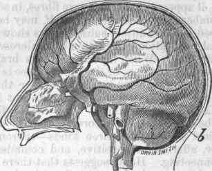
Fig. 8. - The Brain enclosed in the Dura Mater.
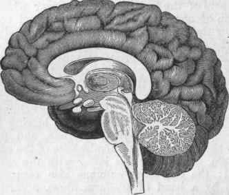
Fig. 4. - Profile view of Human Brain, in vertical section, showing the medulla oblongata, the tuber annulare, the middle portion of the cerebellum with the arbor vitae, the central parts of the cerebrum, and the convolutions on the inner surface of the hemispheres.
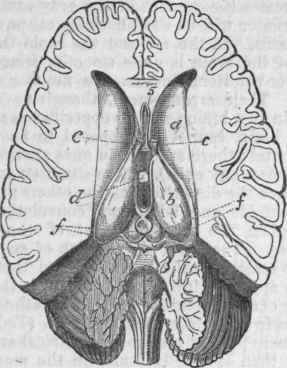
Fig. 5. - Human Brain cut open horizontally, to show its internal parts, a. Corpus striatum of the right side. b. Optic thalamus of the right side. Both these hodies are seen on the floor of the lateral ventricle, which is represented as cut open. c. Anterior pillars of the fornix, cut off at the level of the corpus striatum, d. The middle transverse commissure, passing across from side to side, between the two optic thalami. e. The pineal body. f,f. The tuber-cula quadrigemina. 4. The fourth ventricle, situated just above the medulla oblongata. 5. The fifth ventricle, situated in the substance of the septum lucidum.
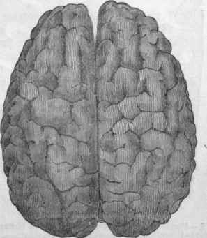
Fig. 6. - Cerebral Hemispheres, viewed from above.
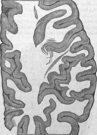
Fig. 7. - Portion of Eight Hemisphere of the Human Brain, divided horizontally, showing the convolutions, and arrangement of white and gray matter.
1, by the blood ascending against gravity;
2, by the curving of the vessels like the letter S before they enter the cranium, thus scattering the force of the stream in different directions; 3, by the minute subdivision of the vessels before they enter the cerebral substance. The impure blood returns through the jugular veins; hence any compression of these vessels by a tight neck stock, or the like, impedes the whole cerebral circulation, causmg, it may be, dangerous congestions. If the blood could be shut off .completely from the brain, death would instantly ensue; and to prevent the possibility of this accident, the vertebral arteries are protected by the bony canals of the cervical transverse vertebral processes from all danger of compression or ordinary injuries. The brains of persons who have died by hanging always exhibit great venous congestion. The veins of the dura mater are quite remarkable by pouring their contents into the large canals enclosed between its layers, the sinuses; these, unlike ordinary veins, cannot be distended beyond a certain point, and, as they all empty their blood into the internal jugular vein, any obstruction in this or in the superior vena cava very speedily produces an uncomfortable distention in the head.
These sinuses are the superior longitudinal, corresponding to the superior margin of the falx cerebri, commencing near the root of the nose (crista galli) and terminating in the cavity called torcular Herophi-1% near the internal occipital protuberance; the inferior longitudinal sinus runs along the lower border of the falx, and ends in the straight sinus, which runs in the median line at the meeting of the falx and the tentorium, and opens into the torcular; the lateral sinuses extend from the torcular downward and forward to the jugular veins. This is the largest sinus, and its canal is deeply hollowed out of the occipital and temporal bones; that of the right side is generally the larger. The sinuses are sometimes the seat of dangerous inflammation. Between the layers of the falx cerebelli are the occipital sinuses, opening into the torcular; the petrosal sinuses, running along the petrous portion of the temporal bone, open into the lateral sinuses; the cavernous sinuses are on each side of the sella turcica, communicating with the petrosal by the transverse sinus, and with each other by the circular sinus.
From this arrangement of the sinuses, communicating freely with the external vessels, may be understood the signal advantages of local depletion in relieving vascular fulness within the head, and also the utility of cold applications for similar purposes. - There are 12 pairs of nerves belonging strictly to the brain, which differ from spinal nerves only in their distribution and in coming through openings in the skull instead of between the vertebrae; all, except the first, proceed from the spinal cord itself, or from its prolongation in the brain (the medulla oblongata). These nerves are: 1, the olfactory, or nerve of smell; 2, the optic, or nerve of vision; 3, motores oculorum, the motor nerves of all the muscles of the orbit, except the superior oblique and the external rectus; 4, thepathetici; 5, the trifacial or trigeminus, the general sensory nerve of the head and face; 6, the abducentes oculorum; 7, the facial, the motor nerve of the face; 8, the auditory, or nerve of hearing; 9, the glossopharyngeal, supplying part of the sensory fibres of the tongue, and presiding over the movements of swallowing; 10, the pneumogastric, or par vagum; 11, the spinal accessory (the last two combined presiding over the functions of respiration and phonation); and 12, the hypoglossal, the motor nerve of the tongue.
Philosophical anatomists have combined these nerves in various ways, separating the three nerves of special sense, and classifying the others in groups resembling spinal nerves, with their anterior motor, and their posterior sensitive roots. As the skull may be considered as composed of three cranial vertebras, we have the olfactory, optic, and auditory special nerves, making their way out through the three vertebra) which may be called by the same name, corresponding to the three primary vesicles which are developed into the brain. Of the intervertebral, analogous to spinal nerves, the first group is composed of the fifth pair for its sensory portion, and of the third, fourth, and sixth for its motor portion; secondly, we have the facial and glossopharyngeal nerves combined; and lastly, the par vagum and spinal accessory form the third group; the hypoglossal may be considered as the first of the true spinal nerves. For further details on this subject the reader is referred to the works of Oarus, Oken, Owen, and other writers on philosophical anatomy. The nature of the nervous force, the functions of the nerves, and the general physiology and pathology of the subject, will be treated under Nervous System; only a brief summary can be given here.
Without question the various operations of the mind are associated with the cerebral convolutions; perception, memory, the power of abstraction, imagination, etc, possess, as instruments of action, the folds of gray matter; as Cuvier says, these parts are the sole receptacles in which the various sensations may be as it were consummated, and become perceptible to the animal. Mechanical injury to the convolutions and the central white substance occasions no pain nor disturbance of the motive powers; in many diseases of the brain and its membranes convulsions accompanied by pain occur, but this depends on a change produced in the striated and optic bodies, and through them propagated to the motor and sensitive nerves. On removing the hemispheres animals are thrown into a state of deep sleep, retaining their muscular power, yet apparently incapable of a single mental nervous action, voluntary or sensory. When the membranes are inflamed, especially the pia mater, the mental faculties are always disturbed; in the delirium of fevers, in delirium tremens, etc, the circulation of the convolutions seems to be disturbed.
The convolutions, then, are the centre of the intellectual actions; being connected with the striated and optic bodies (which have been regarded as the centres of volition and sensation), the intellectual centre may either excite or be excited by them. When the convolutions are insufficiently supplied with blood, the deficient nutrition occasions deranged phenomena of thought and a rapid development of ideas, which, being ill or not at all regulated by the will, assume the forms of delirium and insanity, just as disease of the nerves of vision and hearing may produce unnatural sights and sounds. As in every muscular action some portion of the muscular tissue is wasted, to be supplied by the general nutrition of the body, so every thought is doubtless accompanied by some change in the nervous centre. Concussion of the brain from a fall or blow, or condensation of its substance by a clot of blood, checks the organic changes of the surface, and interrupts the joint actions necessary for consciousness. Gall, the founder of phrenology, assigned to certain convolutions certain faculties of the mind, moral feelings, and instinctive propensities.
This theory has since his time been pursued with the zeal which must naturally attach itself to any science which professes to read the mental tendencies from external signs. In regard to phrenology, it can only be remarked here that, while it is undoubtedly true that the* energy of a nervous centre bears a certain relation to its size, the stress laid by its followers on the temperaments shows that they consider the quality of the brain an important element in the development of nervous power. - During sleep the nervous centres obtain the rest necessary to repair the waste of daily activity; in this state the brain refuses or is slow to convey impressions from without. In deep sleep we are unconscious, and maybe motionless; as the sleep becomes lighter, consciousness begins to return, and mental changes take place, constituting dreams of various kinds. Man performs many actions instinctively, without the intentional adaptation of means to ends, just as the bee makes its cell, or the bird its nest; children are tforn and live for some time without cerebral hemispheres, who perform the acts of sucking and swallowing perfectly well; remove the hemispheres in an animal, and it will eat if food be placed in the mouth, though it will not go to seek it; many idiots will do the same.
In what part of the brain resides the power presiding over these actions? At the base of the brain, concealed by the hemispheres, is a series of ganglia, the origin of the nerves of special sense, as well as the striated and optic bodies into which all the fibres connecting the hemispheres with the medulla oblongata pass; these nerves have therefore their own nervous centres, distinct in function from other parts of the brain. In fishes these ganglia are very large, and the hemispheres comparatively small, sometimes smaller than a single pair, the optic. In man the instinctive propensities are in a measure superseded by intelligence, but they may act independently of it. The real nervous centres for motion and sensation are those situated at the base of the brain, and not the hemispheres; as far as mere animal life and motion are concerned, the latter are not essential; a vast proportion of animated creatures (all the invertebrata) have no trace of them; they are added in man for the intellectual and moral nature.
The instinctive and emotional actions are excited through the special ganglia, following directly upon sensation, without any process of thought; they are sometimes stronger than the voluntary actions; e. g., we are often compelled to laugh at something ludicrous though we have the strongest motives not to do so. Long-continued habit will often make us perform actions instinctively, as it were, which at first required an effort of the will: for instance, in an old snuff-taker, who had been seized with epilepsy, irritation of the nose with a feather to restore consciousness produced a contraction of the right forefinger and thumb to take a pinch. These emotional actions may be excited by mental operations. Whenever the feelings get the better of the reason, the sensory ganglia are excited at the expense of the hemispheres, and the individual is for the time being morally insane, even though these emotions may point in the right direction; fanatics of all classes, in this way, are really insane, generally monomaniacs. These instincts may also be in opposition to the reason, and then the more a man follows them the closer does he approach the brutes.
Comparative anatomy teaches that the cerebellum is largest in those animals which have the greatest variety of motions; injury or removal of this organ causes no pain nor convulsions, but destroys the power of combining properly the voluntary motions. Man, though inferior to many animals in particular kinds of movements, far surpasses them in the number and complexity of their combinations; the act of walking brings into action almost every muscle of the trunk and extremities, and is superior to all other modes of exercise. In man the cerebellum attains its highest development. Inflammation of its membranes, and even its almost complete destruction by slow disease, has little effect on the intelligence, but the motive powers are disturbed. In intoxication the energy of the cerebellum is first destroyed, and afterward the intelligence and consciousness, leaving the subject for the time little better than dead, motionless, and insensible. The distinct operation of these various centres is made obvious by many conditions of the body, in which one or more are inactive.
In deep sleep, the hemispheres, the sensory ganglia, and the cerebellum are more or less completely at rest, but the medulla oblongata and the spinal cord must, as always, be wide awake; in dreaming, the hemispheres are partially active; in somnambulism, a step nearer to wakefulness, the hemispheres are awake, and also the cerebellum, so that the movements are well adapted to the thoughts. It is well known that in this state persons have walked over dangerous places which they could never have passed in open day; there is an evident loss of control over the thoughts, which are more influenced by external impressions than in dreaming, so that the somnambulist may answer questions properly; that there is not full command over the senses, the dangerous accidents occurring in this condition fully prove; the events of this state may not be remembered in the waking hours, but may be taken up again by the memory the next night, constituting complete "double consciousness." A condition remarkably analogous to somnambulism is the mesmeric sleep or trance; a nervous habit of body predisposes to both.
Overworking the brain exhausts the body; wear and tear of the brain, like wear and tear of the muscles, require periodic and long intervals of rest; from want of attention to this fact, many a bright intellect has faded into imbecility and insanity. - The primary ganglia of the vertebrate brain are three in number, and they are developed into the anterior or cerebrum, the posterior or cerebellum, and the median or quadrigeminal bodies. In fishes, the lowest vertebrates, the medulla is large, with the pyramidal and restiform bodies, but without the olivary; the brain looks like a series of ganglia developed on the superior surface of the cord, two pairs and a single one: 1, the olfactory lobes, analogous to the hemispheres in man, from which the nerves of smell arise; 2, behind these, the optic lobes, generally considered analogous to the tubercula quadrigem-ina, in some fishes larger than the other parts of the brain; from these arise the optic nerves, and the third, fourth, and sixth pairs; 3, behind these, the imperfectly developed cerebellum generally, but of large size in the selachians.
In reptiles the brain well fills the cranial cavity, and the preponderance of the spinal cord is less; the olfactory lobes, now obviously the hemispheres, are increased in size, with an internal cavity, and a commissure; the second cerebral mass and its cavities are smaller; the cerebellum is small in the lower orders, but with lateral appendages and external striae in the higher. In birds the brain and spinal cord are no longer on the same plane; the brain is the larger, and the ganglia are more above and less behind each other; the hemispheres are larger than the other parts, are united by commissures, and contain true lateral ventricles in which is a tubercle resembling a corpus striatum; the optic lobes are small, separated, with smaller cavities; the cerebellum is particularly large, with evident lateral lobes and external striae. In mammals the brain is much larger than the cord; the cerebral hemispheres are of large size, with marked convolutions in the higher orders, with a corpus callosum, lateral ventricles with anterior, descending, and (in the monkey) posterior horns, optic and striated bodies, taenia semicircularis, and fornix; the optic lobes are small, divided into two pairs, solid, and are now called the tubercula quadrigemina; the cerebellum is highly developed, the more so as the animal approaches man, presenting the arbor vitae in its interior; the pons Varolii is large, and the fourth ventricle is completely concealed and . shut in.
Prof. Owen has divided the mammalia into four groups, according to. the characters of the surfaces of the cerebral hemispheres; in some the hemispheres are but feebly connected by the fornix and anterior commissure, while in the great majority the corpus callosum is added; in the former case there is a peculiar mode of development of the young owing to the absence of the placenta. His groups are as follows: 1, lyencephala, having the hemispheres loose and disconnected, leaving exposed the olfactory ganglia, cerebellum, and more or less of the optic lobes, their surface smooth, or with very few anfractuosi-ties; this includes the marsupials; 2, lissence-phala, having a corpus callosum, with the cerebellum and olfactory lobes exposed, the surface smooth, or with very few and simple convolu-. tions; this includes the rodents, insectivora, cheiroptera, and edentata; 3, gyrencephala, having the cerebrum extending over more or less of the cerebellum and of the olfactory lobes, with more or less numerous convolutions; this includes cetacea, pachydermata, herbivora, carnivora, and quadrumana; 4, archencephala, embracing man only. - Those wishing to pursue the study of the brain are referred to the works of Solly, Longet, Leuret, Todd and Bowman, Carpenter, Owen, Tiede-mann, Mtiller, and to the researches of J. Lock-hart Clarke on the " Intimate Structure of the Brain, Human and Comparative," and those of Dr. John Dean "On the Gray Substance of the Medulla Oblongata and Trapezium".
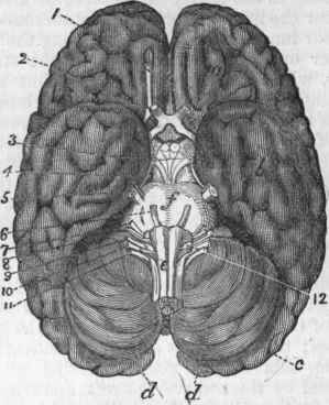
Fig. 8. - Human Brain, viewed from below, a. Anterior lobe of cerebrum, b. Middle lobe of cerebrum, c. Posterior lobe of cerebrum, d. Cerebellum, e. Medulla oblongata, f. Tuber annulare. 1. Olfactory nerves. 2. Optic nerves. 3. Motores oculorum. 4. Patbetici. 5. Trifacial. 6. Abducentes oculorum. 7. Facial. 8. Auditory. 9. Glossopharyngeal. 10. Pneumogastric. 11. Spinal accessory. 12. Hypoglossal.
Continue to:
My Books

