Callosities (Chestnuts And Ergots). Part 3
Description
This section is from the book "The Horse - Its Treatment In Health And Disease", by J. Wortley Axe. Also available from Amazon: The Horse. Its Treatment In Health And Disease.
Callosities (Chestnuts And Ergots). Part 3
Among asses, chestnuts are usually found in the distinctly modified form described - i.e. bare patches of skin, often rather larger and more circular in form than the chestnuts of the horse, - and to the naked eye are covered with thickened epidermis. It may be added, however, that in some specimens of chestnuts recently obtained from asses the horny substances projected something like ¼ of an inch above the surface of the skin, in fact they were larger than some which have been lately obtained from the legs of well-bred horses.
In the following illustrations (fig. 665) are represented a chestnut from the fore-leg of a cart mare and one of the ergots from the fetlock joint: also specimens of a chestnut or bare patch from the fore-leg of an ass and one from the fore-leg of a foetus of a mare at about the eighth month of gestation.
To the naked eye the chestnuts of the ass and those of the foetus of the mare are identical in appearance, differing altogether from the chestnuts of the adult horse; but under the microscope the three forms are seen to be essentially the same in their minute structure (Plate LXIX).
That all the cuticular appendages, hair, nail, and horn, are composed of epidermic cells arranged in various ways is quite well known. To assert, therefore, in respect to any of the structures, that they are hardened, condensed, or modified cuticle is correct; at the same time the statement is not sufficiently definite from the point of view of the scientific enquirer.
Cuticle or epidermis is arranged in the manner of the tiles or the slates on the roof of a building, each cell representing a tile.
Horn is formed by the secretion of cells round a cone or villus projecting from the underlying vascular membrane, and assumes in consequence the form of hollow fibres closely felted together.
Hair is developed in a similar manner from a papilla at the bottom of a small depression or follicle, the chief difference being that each of the hairs is distinct. They are not felted together or arranged in masses, as in horn.
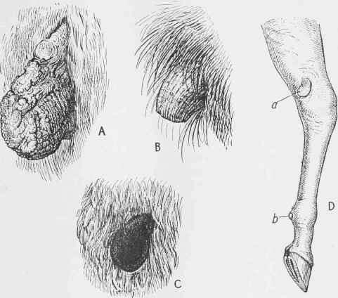
Fig. 665. - A, A large chestnut from a cart mare. B, Ergot from same animal. C, Bare patch from fore-leg* of an ass. D, Bare patch from foetus of mare. All about 2/3 of natural scale. a, Chestnut; b, Ergot.
Nail is also formed from a villous membrane, the fibres being very fine, and densely crowded together, constituting an extremely hard structure.
Although hoof, hair, and nail are all composed of the same elements, the difference in their arrangement is so distinctive that a tyro in the use of the microscope finds it a perfectly easy task to recognize and to name the several structures when placed before him.
The present enquiry is to ascertain the structure of those peculiar formations on the legs of the horse family, known as chestnuts, ergots, and bare patches of cuticle, and also of the parts described as plantar and palmar pads in man and certain animals, with the view to placing them in the classes of substance to which they respectively belong.
Perhaps the most simple way of performing the task will be to classify the several structures at the commencement, and then to show by description and illustration how the classification has been arrived at.
Proceeding on this plan, the structure classed as horn will include all the growths known as chestnuts, ergots, and bare patches of hardened cuticle, notwithstanding the decided variations of form which they present to the naked eye.
In the next class - "Modified epidermic covering" - must be placed all the varieties of plantar and palmar pads.
Taking the chestnuts or ergots of the horse first, both in the adult and in the foetus near the time of birth, there is no difficulty in showing that they are horny structures. The sections, both transverse and vertical, exhibited in the following diagrams (fig. 666) and in Plate LXIX place the matter beyond doubt, and it will be interesting to compare the different sections with the objects as they appear to the naked eye in fig. 665, a, b, C, D. The bare patches covered with hardened cuticle in the ass, and the similar bare patches in the foetus of the mare, are, as previously stated, both quite distinctly true horny structures developed from a villous membrane, exactly as the perfectly formed horny excrescences (chestnuts) are in the adult.
After maceration in water in the case of the chestnuts of the ass, and without any preparation in those of the foetal horse, or of the foal at birth, the epidermic covering may be stripped off, and with a pocket lens the secreting membrane thus exposed may be seen covered with villi. The thin layer which has been stripped from it may by the same instrument be resolved into a fine plate of horn identical with the horn of the sole. Transverse and vertical sections under the microscope show all the details of the structures, the sudden transition from ordinary skin to the villous secreting membrane and the horny covering on the surface. All these parts are indicated in the figures referred to, which may be taken to represent the minute structure of the organs exhibited in fig. 665, A, B, c, D.
Sections of the ergot in the horse and in the ass exhibit exactly the same intimate structure. In fact they differ only in the size and form of the horny growth. No further proof can be required in support of the statement that chestnuts and ergots, whether they appear as horny excrescences or as bare patches of hardened cuticle, are in their minute structure identical with hoof horn, and further, that the membrane from which they are developed is a vascular villous membrane, precisely analogous to the villous membrane of the coronary surface and sole of the horse's foot, which has already been described and figured on pp. 434 and 435 of this volume.
These facts would have consisted remarkably well with the theory of their being remnants of digits, were it not for the cogent objections which have been urged against that view. As it is, the identity of structure in the horny growths and the horn of the foot does not tend to assist in the attempt to assign to them any special economy, or in any way to indicate what functions they might have possessed in their more developed condition.
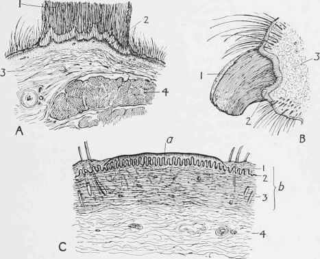
Fig. 666. - Sections of Chestnut and Ergot of Horse and Bare Patch of Ass.
A, Horizontal section through chestnut of horse - 1, horn; 2, villous secreting membrane; 3, subcutaneous tissue; 4, muscle. B, Perpendicular section through ergot of horse - 1, horn; 2, villous secreting membrane; 3, subcutaneous tissue. c, Section through bald patch of ass: a, the bare patch; b, skin - 1, horny layer of epidermis; 2, malpiguian (mucous) layer of epidermis ; 3, derma; 4, subcutaneous tissue.
Plantar pads are represented in fig. 664, A, B (p. 503) in man and dog, and their corresponding positions in the leg and foot of the horse are indicated at c in the same figure. It has been stated already that these pads in man are really hardened cuticle, excessively thick portions of the cuticle in fact.
In the foot of the dog, however, both on the surface and in section, the structure differs from the thickened cuticle of man's hand and foot, and also from true horn. Indeed, the minute anatomy of the organ exhibits a most perfect type of the transition or change from cuticle to horn. (See Plate LXIX.)'
In the case of the dog the plantar and palmar pads are in perfect form and active function. In man, however, they are more or less accidental or rudimentary. The palmar pads, or those on the palm of the hand, depend for their development on the amount of manual work done, and they vanish when that work ceases, while the growth of the plantar pads is checked by the devices of civilization, including shoes and stockings, and the use of various modes of locomotion in place of the natural acts of running and walking.
Continue to:
My Books

