The Development Of The Embryo. First Signs
Description
This section is from the book "The Horse - Its Treatment In Health And Disease", by J. Wortley Axe. Also available from Amazon: The Horse. Its Treatment In Health And Disease.
The Development Of The Embryo. First Signs
At the outset, the attempt to describe the formation of the various parts of the young animal is met by an insuperable difficulty, because by no form of verbal gymnastics is it possible to describe a whole set of simultaneous processes by the aid of consecutive phrases. It is easy, for example, to state the fact that in the germinal membrane the embryo is formed; that bones, muscles, integument, and viscera appear, and that adaptive changes go on in the uterus, in which the young one has to pass its embryonic and foetal life; but unless the reader will consent to make a mental effort to realize that the changes are all going on in different degrees at the same time, there is no hope that the writer will succeed in conveying a correct idea of the true nature of the developmental process.
Proceeding from the point which has just been reached, the formation of a germinal membrane by an accumulation of cells round the inside of the investing membrane of the yolk (yolk-sac), it will be quite easy to understand that at a certain part in the blastodermic membrane a round mass of cells appears, called for the sake of distinction the germinal area. In this round mass, which soon becomes an oval mass, the first sign of the embryo is seen, as shown in the accompanying figure (fig. 538, e).
On each side the primitive groove or trace above described, are collected two oval masses of cells rising above the plane of the germinal membrane and bending towards each other until they touch and form an arch in which the incipient spinal cord is to be lodged; all this is arranged, it must be observed, in the upper or serous layer of the germinal membrane. Immediately below the primitive groove a line of cells may be recognized, forming the chorda dorsalis, the rudimentary stage of the bodies of the bones of the back (dorsal vertebrae). Then below the primitive groove, at the same time that the cells of the laminae dorsalis are closing over to form the central canal for the spinal cord, the serous membrane sends off prolongations from its lower margin, the laminae ventrales, which unite to form the walls of the trunk to enclose the abdominal viscera.
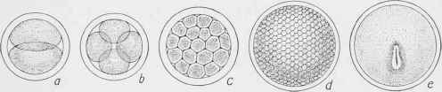
Fig. 538. - The Development of the Ovum a, First division of the ovum; b, c, d, subdivision of the ovum; e, first trace of the embryo.
As they proceed downwards, the ventral lamina turn inwards, enclosing part of the yolk-sac, after which the yolk and inner mucous layer of the germinal membrane are divided into two portions, one being retained in the body of the embryo, the other being left outside. The latter is called the umbilical vesicle. The mucous layer of the germinal membrane now lines the interior of the abdominal cavity and also the interior of the umbilical vesicle. The upper or serous layer is continued round both, and from the portion of the mucous layer enclosed in the body of the embryo the intestinal canal is developed.
This state of the embryo is represented in the next illustration (fig. 539).
Continue to:
My Books

