Foetal Membranes
Description
This section is from the book "The Horse - Its Treatment In Health And Disease", by J. Wortley Axe. Also available from Amazon: The Horse. Its Treatment In Health And Disease.
Foetal Membranes
While the changes above described have been going on, the formation of the foetal membranes, the allantois and amnion, is proceeding. Folds of the external layer of the blastodermic membrane are raised to enclose the body of the embryo forming the amnion; at the same time during the development of the amnion the allantois protrudes from the hinder portion of the intestinal canal, as a small pear-shaped mass of cells at first, but, rapidly extending, it presses its way between the folds of the amnion and comes in close contact with the outer one of the two folds, becoming more vascular as it proceeds. Reaching the umbilicus, the allan-tois is divided into two parts. The outer part, however, extending to the external investure of the ovum, the chorion, shrivels, and is lost; the other portion remains in the abdominal cavity, and part of it is converted into the urinary bladder, while the remaining portion extends from the bladder to the umbilicus under the name of urachus, which after birth forms one of the ligaments of the bladder.
It may be remarked here that an oval body flattened in form, which is commonly described as a false tongue, and sometimes affirmed to exist in the mouth of the foal, is really a concretion which is met with in the fluid of the allantoid sac, and nowhere else; occasionally there are several of these bodies, of various sizes. The name given to them, "Hip-pomanes", indicates that they were known to the Greeks, and an ancient superstition attributed to them talismanic power, a belief in which still exists in some parts of the country.
The annexed figure (fig. 539) shows the arrangement of the three membranes which invest the ovum, i.e. the external chorion, the amnion, the outer portion of which becomes in part firmly attached to the inside of the chorion, and the allantoid sac.
The villi on the outer surface of the chorion of the human ovum (fig. 540) are seen to be massed on the right side of the figure to form the placenta. In the equine ovum there is no circumscribed placenta, but instead the vascular villi are connected throughout with the internal uterine membrane by means of numerous placental tufts, which penetrate the lining of the uterus so that the capillaries of the foetal vessels and those of the maternal vessels are in contact over the whole surface. There is, however, no actual communication between the two sets of capillaries, but the bloodstream of the mother and that of the foetus are separated only by the thin walls of the vessels, through which the blood is constantly flowing. The interchange which takes place between the maternal and the foetal blood, for the nutrition of the young animal, necessarily is carried on through the two layers of membrane by osmosis, i.e. that force which regulates the interchange of fluids through wet membranes.
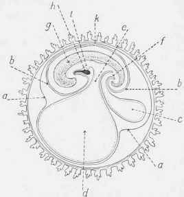
Fig. 539. - Development of the Embryo, eighteenth day.
a, Outer or corneous layer; b, amnion; c, allantois connected with the anal portion of the alimentary canal; d, yolk-sac or umbilical vesicle ; e, vitello-intestinal opening; f, simple alimentary canal in lower position; g, trunk and head of embryo; A, foetal heart; i, alimentary canal in upper portion; k, place of convergence of amnion and reflexion of false amnion or corneous layer.
Blood-vessels in the embryo commence by formation of a thin membrane in the blastoderm, between the serous and mucous layers, at a part which is described as the vascular area. Red lines appear, and form a network of vessels filled with blood, a rudimentary heart is formed in the vascular area, and to that organ the branching vessels proceed, and the outline of the circulatory system is complete; the details being filled in by further developments in correspondence with the continuous advance of the embryonic structures.
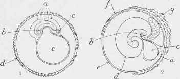
Fig. 540. - Development of the Human Ovum.
1. Early stage: a, interior and exterior folds of the serous layer joining the amnion; 6, embryo; c, incipient allantois; d, chorion; e, vitelline mass surrounded by blastodermic vesicle. 2. Second month: a, amnion, outer layer, coalescing with chorion; b, embryo; c, umbilical vesicle; d, amnion, inner layer; e, smooth portion of chorion; f, villous portion of chorion; g, elongated villi collecting into placenta.
In the next illustration (fig. 541) the condition of the embryo and its membrane at the age of seven weeks is shown.
Continue to:
My Books

