


Clinical And Experimental Studies On Serum Pyridine Nucleotide. Part 2
Description
This section is from the book "The Scientific Contributions Of The Ben May Laboratory For Cancer Research", by The University of Chicago. Also available from Amazon: The Scientific Contributions Of The Ben May Laboratory For Cancer Research.
Clinical And Experimental Studies On Serum Pyridine Nucleotide. Part 2
In portal cirrhosis, serum ICD was usually within the normal range and was never elevated to values greater than three times the normal mean, even in paiients in hepatic coma. In cases of extrahepatic obstruction of the common bile duct, resulting from either benign or malignant causes, serum ICD levels fell invariably within the normal range. In about 50 per cent of patients with objective evidence of liver metastases from malignant disease, the serum ICD activities were greater than the upper limit of the normal. In these situations, however, serum ICD was never increased more than six times the normal mean, in marked contrast to the much larger elevations in the activity of this enzyme which were often observed in viral hepatitis. A wide variety of malignant tumors did not cause elevations of serum ICD. If serum ICD is elevated in malignancy, involvement of the liver is indicated.
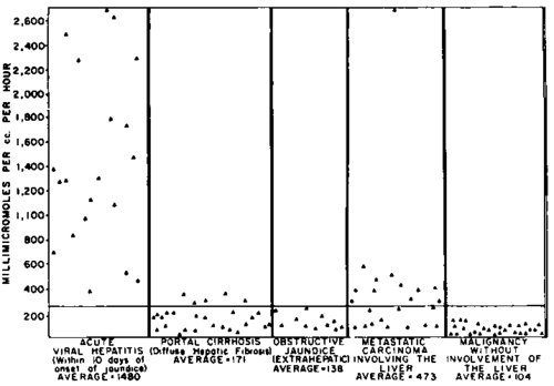
Figure 1. Serum isocitric dehydrogenase levels in human liver disease. The horizontal line at the level of 264 represents the upper limit of normal activity for scrum ICD.
Serial estimations of serum ICD, GPT, and LD activities in human liver disease" showed that alterations in GPT activity were somewhat similar to those of ICD, whereas the changes in LD followed a different pattern and were always of a smaller magnitude than those of ICD and GPT. In viral hepatitis, elevations of serum GPT were sometimes of greater magnitude than those of serum ICD. In patients with the usual benign course of this disease, serum GPT levels tended to remain increased for longer periods of time than those of serum ICD. Moreover, in extrahepatic obstruction, serum ICD activities were never abnormal, whereas those of serum GPT were occasionally elevated, as others27 have noted. Changes in serum ICD in liver disease bore no relationship to parallel alterations in serum alkaline phosphatase activity and could not be correlated with the extent of hyperbilirubinemia or with the results of flocculation tests.
Serum 6-phosphogluconic dehydrogenase in disease. Studies on a more limited number of patients than were examined for serum ICD activity showed (Wolf-son and Williams-Ashman, unpublished data) that serum PGD levels were seldom abnormal in the absence of liver disease. Normal values for PGD were found in seven patients with carcinoma of the breast with extensive osseous metastases and in five patients with carcinoma of the prostate. Serum PGD levels were not increased in cirrhotics. Elevations in serum PGD were observed during the acute phase of viral hepatitis and occasionally in patients with malignant disease with metastases to the liver. The changes in serum PGD in liver disease were never as great as parallel alterations in serum ICD.
Alcohol and polyol dehydrogenase activity of serum. Mammalian liver is known to contain a DPN-linked enzyme, alcohol dehydrogenase (AD), which catalyzes the oxidation of ethanol to acetaldehyde:88
CH3CH2OH + DPN+ ⇋CH3CHO + DPNH + H+
Another distinct enzyme oxidizes a number of 5-, 6-, and 7-carbon polyols to the corresponding ketose sugars, for example:
Sorbitol + DPN+ ⇋ D-Fructose + DPNH + H+
L-Iditol + DPN+ ⇋ L-Sorbose + DPNH + H+
The activity of this enzyme, polyol dehydrogenase (PD) is greatest in liver and kidney.89 30 Neither of these enzymes could be detected in normal human blood serum, as evidenced by the oxidation of DPNH by either acetaldehyde or d-fructose. However, marked activity of both enzymes was found in the sera of certain patients with acute viral hepatitis, or with liver metastases from malignant disease. A typical spectrophotometric determination of these enzymes in the serum of a patient with acute viral hepatitis is depicted in figure 2. In these assay systems, the rate of oxidation of DPNH by either acetaldehyde of D-fructose was found to be strictly proportional to the amount of serum added. It will be seen from table 1 that the activities of AD and PD in serum bore no constant relationship to one another in patients with viral hepatitis exhibiting the typically increased values of serum ICD. Since neither AD nor PD are present in normal blood serum, the finding of these enzymes in serum is a qualitative systemic index of liver disease.
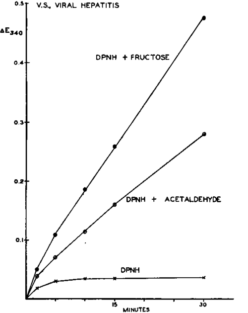
Figure 2. Alcohol and polyol dehydrogenase activity of the serum of a patient with acute viral hepatitis. Each vessel contained 100 µmole Tris(hydroxymethyl)aminomethane buffer of pH 7.4; 150 µmole NaCl and 0.3 µmole DPNH in a final volume of 3.0 ml. If added: acetaldehyde 20µmole; D-fructose 50 µmole. Change in optical density at 340 mµ plotted from the time of addition of serum (0.5 ml.) to the reaction mixture. Temperature 25° C.
Fate of injected isocitric dehydrogenase. ICD was purified from rat heart by a slight modification of the method of Siebert et al.31 The purified material catalyzed the oxidation of approximately 2000 mumoles of isocitrate per milligram of protein per minute under the conditions of the ICD assay system.17 The preparation was devoid of glutamic dehydrogenase activity, as evidenced by its inability to catalyze the oxidation of DPNH upon the addition of a-keto-glutarate and ammonium chloride at pH 7.4. Just before administration to experimental animals, the purified enzyme was dialyzed thoroughly against 0.15 M NaCl at 2° C. The enzyme was injected into the saphenous vein of rats weighing approximately 300 gm. under ether anesthesia. Sufficient amounts of enzyme were injected to raise the serum ICD activities thirtyfold to seventyfold within 2 minutes after injection. Blood samples were removed at various time intervals from the jugular vein. From semilogarithmic plots of serum ICD activity versus time, the mean half life of the injected enzyme was found to be 60 min. No difference in the rate of disappearance of injected ICD from the blood stream was observed between male and female animals of the same age. Rat blood serum incubated with purified ICD at 37° C. inactivated the enzyme at very slow rates; more than 50 per cent of the activity remained after 24 hours.
Table 1. Alcohol And Polyol Dehydrogenase Activity Of The Sera Of Patients With Viral Hepatitis
Patient | Enzyme activity* | ||
Isocitric dehydrogenase | Alcohol dehydrogenase | Polyol dehydrogenase | |
1 | 3400 | 493 | 384 |
2 | 768 | 133 | 12 |
3 | 390 | 134 | 260 |
4 | 3480 | 445 | 778 |
5 | 4500 | 822 | 480 |
• Mimoles substrate oxidized or reduced per 1 ml. serum per hour at 25°C. Isocitric dehydrogenase determined by the method of Wolfson and Williams-Ashman.17 Alcohol and polyol dehydrogenases determined under the conditions shown in figure 2.
The rapid disappearance of activity after the injection of enzymes directly into the circulation has been noted by Warburg' for aldolase and by Huggins*2 for acid phosphatase. It would appear that the maintenance of greatly increased serum ICD levels following acute injury to previously healthy hepatic cells (as, for instance, in acute hepatitis) must entail a continual outflow of this enzyme from dead or damaged cells into the blood stream.
Carbon tetrachloride poisoning. Soon after the administration of carbon tetrachloride, the liver parenchyma undergoes an inflammatory reaction which is usually followed by massive necrosis. The hepatotoxic action of carbon tetrachloride appears to be related to a disruption of mitochondrial structure by this agent.33 The hepatic lesions produced by carbon tetrachloride bear considerable resemblance to those resulting from hepatitis of viral origin. Large increases in the activity of some, but not all, enzymes in blood serum have been reported to follow the administration of carbon tetrachloride to experimental animals. For instance, the careful studies of Bruns and Neu-haus34 demonstrated that the aldolase and phosphohexoseisomerase activity of mouse serum rose enormously in animals poisoned by carbon tetrachloride, while the levels of these enzymes in liver fell dramatically; under the same experimental conditions, serum tributyrinase, amylase, and alkaline phosphatase were barely affected. In order to gain some insight into the factors which govern the efflux of enzymes into the circulation following liver damage, the serum levels of ICD, PGD, LD, AD, and PD were studied at various time intervals after administration of carbon tetrachloride. All of these enzymes appear to be confined mainly to the soluble fraction of liver cells, that is, they are not bound to either mitochondrial or microsomal structures, and all of them use a pyridine nucleotide as a coenzyme. The carbon tetrachloride was dissolved in mineral oil and given in a single dose (5 cc. CCl4/kg.) by stomach tube to Sprague-Dawley rats weighing between 350 and 400 gm. Blood samples were taken from the abdominal aorta under ether anesthesia. It will be seen from figure 3 that serum ICD activity rose as much as four hundredfold after the injection of this poison. In the same animals, marked AD and PD activity appeared in the sera of the animals which had received carbon tetrachloride. The change in activity of all of these enzymes occurred more rapidly in female than in male rats of the same age. Serum PGD levels increased after the administration of carbon tetrachloride, but to a much lesser extent than those of serum ICD; serum LD was altered only to a very small extent.
Continue to:


My Books

