


VI. Fate Of Steroid Estrogens In Target Tissues
Description
This section is from the book "The Scientific Contributions Of The Ben May Laboratory For Cancer Research", by The University of Chicago. Also available from Amazon: The Scientific Contributions Of The Ben May Laboratory For Cancer Research.
VI. Fate Of Steroid Estrogens In Target Tissues
Elucidation of the process by which the steroid sex hormones exert their remarkable stimulation of the growth of certain mammalian tissues is one of the major problems of current interest to biological scientists. Consideration of the mechanism of steroid hormone action is seriously handicapped by lack of knowledge of the chemical transformations which the steroid molecule undergoes during the growth-induction process. Although a large number of metabolic conversion products of steroid sex hormones have been isolated from urine, it has not been established whether any of these substances are produced in the target tissues when they are stimulated to grow. As an approach to the understanding of estrogen action, an attempt has been made to determine the fate of physiological amounts of estradiol, as this hormone promotes growth of the uterus and other responsive organs of the immature rat.
In the case of the estrogens, the problem is complicated by the fact that minute doses of steroids are effective in eliciting the biological response. It is necessary to work within the physiological dose range if the fate of the steroid in the target tissues is to have relevance to growth promotion rather than to the disposal of excess hormone by the organism. Special techniques have been developed for the present study. These include an accurate procedure for the determination of tritium in mammalian tissues (III) and methods for the synthesis and manipulation of tritiated steroids of unusually high specific activity. Catalytic reduction of 6-dehydroestradiol was carried out with carrier-free tritium gas in an apparatus developed especially for this purpose (119). The 6,7-tritiated estradiol isolated had a specific activity of 30 to 50 curies per millimole, which permits the accurate measurement of as little as 1 picogram (10^-12 gm.) of the steroid.
With these tools an investigation (119) was undertaken to determine what fraction of an administered physiological dose of estradiol reaches the target tissues, how long it remains there, where within the tissues and within the cells it becomes localized, and what happens chemically to the steroid as it induces growth.
Incorporation Of Steroid In The Tissues
After a single subcutaneous injection of 0.1 µg. of estradiol in saline to the immature female rat, it was found that in blood and most tissues studied the level of radioactivity reaches a maximum within 15 minutes, after which it declines rapidly (Fig. 21). In the uterus and vagina, on the other hand, as well as in the pituitary, the maximum activity is not observed until about one hour after administration, and these tissues show the ability to incorporate and retain radioactive steroid at a time when the radioactivity in the blood and other tissues has fallen to a rather low level. The concentration of radioactivity, especially after one hour, is somewhat greater in the accessory sexual tissues than in the other tissues, but, even so, it represents an extremely small fraction of the administered steroid. The maximum activity reached in the entire uterus corresponds to only 0.10-0.15 per cent of the administered dose, or about 100-150 picograms of steroid.
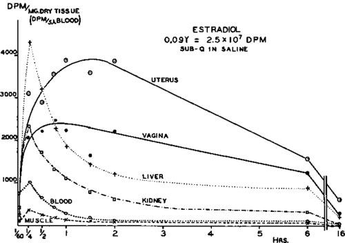
Fig. 21.-Concentration of radioactivity in rat tissues after single subcutaneous injection of 0.098 ng. (11.5 µc.) of estradiol in saline. Liver and kidney points are mean values of 3 aliquots of dried pooled tissue; other points are median values of individual samples from six animals.
When the administered dose of estradiol was reduced to o.oi /xg., incorporation patterns similar to those shown in Figure 21 were observed, although the concentration of radioactivity in the tissues was reduced proportionately.
Continue to:


My Books

